Download
1 / 34
340 likes | 449 Vues
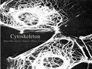
Stable spatial gradients of cytoskeleton assembly regulators. David Odde University of Minnesota. Microtubule Structure. “Catastrophe”. Length (µm). “Rescue”. Time (minutes). Microtubule “Dynamic Instability” (DI). k c. V g. V s. k r. see VanBuren et al., PNAS USA (2002).

E N D
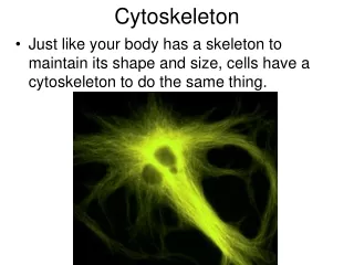
Stable spatial gradients of cytoskeleton assembly regulators David Odde University of Minnesota
“Catastrophe” Length (µm) “Rescue” Time (minutes) Microtubule “Dynamic Instability” (DI) kc Vg Vs kr see VanBuren et al., PNAS USA (2002)
In animal cells: In yeast: 10-20 µm 1.5 µm ~1000 MTs ~40 MTs Mitotic Spindle Interpolar microtubule kinetochore spindle pole body spindle pole body kinetochoremicrotubule chromosome
Hypothesis Dynamic instability alone is sufficient to explain the observed MT length distribution in the yeast mitotic spindle
Results: Cse4p-GFP Distribution ? 2 µm Experimentally Observed Theoretically Predicted
“Catastrophe” Length (µm) “Rescue” Time (minutes) Microtubule “Dynamic Instability” (DI) kc Vg Vs kr
-0.4 -0.2 0 +0.4 μm +0.2 Point Spread Function (PSF) • A point source of light is spread via diffraction through a circular aperture • Modeling needs to account for PSF
Model-Convolution Original Fluorophore Distribution Simulated Image Obtained by Convolution of PSF and GWN with Original Distribution
Results:Distribution of Cse4-GFP fluorescence Experimentally Observed Theoretically Predicted
QS SE QS x=0 x=L Results: Distribution of Cse4-GFP fluorescence
1000 nm Results: DI Only Model
k k* Surface reaction B-->A Homogeneous reaction A-->B MT Repellant Concentration MT Attractant X=L X=0 Position Microtubule Chemotaxis A: Phosphorylated Protein Stabilizes MTs B: Unphosphorylated Protein Destabilizes MTs Microtubule Immobile Kinase Mobile Phosphatase
Microtubule Chemotaxis:Op18 A: Op18-hi-P B: Op18-low-P Destabilizes MTs Chromatin Microtubule Immobile Plx1 Mobile PP2A Op18-low-P Concentration Op18-hi-P Position
Microtubule Chemotaxis: RanGTP A: RanGTP Stabilizes MTs B: RanGDP Chromatin Microtubule Immobile RCC1 Mobile RanGAP RanGDP Concentration RanGTP Position
Model for Chemotactic Gradients of Phosphoprotein State Fick’s Second Law with First-Order Homogeneous Reaction (A->B) B.C. 1: Surface reaction at x=0 (B->A) B.C. 2: No net flux at x=L Conservation of phosphoprotein
Predicted Concentration Profile If k= 1 s-1, D=10-11 m2/s, and L=10 µm, then g=3
Microtubule Chemotaxis: RanGTP A: RanGTP Stabilizes MTs B: RanGDP Chromatin Microtubule Immobile RCC1 Mobile RanGAP RanGDP Concentration RanGTP Position
1000 nm Results: Chemical Gradient and Polar Ejection Force Models
Figure 2 Right Half Spindle Left Half Spindle Cse4 Bleach @ end of simulation, mutant “Tension” model
Figure 4 Right Half Spindle Left Half Spindle Cse4 Bleach @ End of Simulation, wild-type, “Gradient-Only” Model
F F F F Mitotic Spindle Conclusion: Spatial gradients in MT DI parameter(s) may play a role in mediating budding yeast mitotis
Simulated Actin Filament Dendritic Branching Simulated Image of Actin Filament Dendritic Branching Y Y X Z X X Model-Convolution: Application to Dendritic Actin Filament Branching
Original Fluorophore Distribution Simulated Image Obtained by Model-Convolution of Original Distribution Image Obtained by Deconvolution of Simulated Image Potential Pitfalls of Deconvolution
Acknowledgements • Whitaker Foundation • National Science Foundation
Comparing Models to Microscopy Molecular Theory Molecular Reality Computer Simulation Fluorescence Microscope Model Predictions Microscopic Observations ???