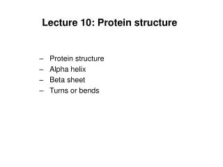
Lecture 10: Protein structure
Lecture 10: Protein structure. Protein structure Alpha helix Beta sheet Turns or bends. Ramachandran Diagrams. Show the allowed conformations of polypeptides.

Lecture 10: Protein structure
E N D
Presentation Transcript
Lecture 10: Protein structure • Protein structure • Alpha helix • Beta sheet • Turns or bends
Ramachandran Diagrams • Show the allowed conformations of polypeptides. • These work, because the sterically allowed values of and can be determined by calculating the distances between the atoms at all values and for the central peptide unit. • Sterically forbidden conformations are those in which any nonbonding interatomic distance is less than its corresponding van der Waals distance. • This info can be summarized by the Ramachandran Diagram or Conformation map
Structural properties predicted by Ramachandran Diagram Regions of “normally allowed” torsion angles are shown in blue. Green regions are the “outer limit” regions. Page 222
Table 8-1 van der Waals Distances for Interatomic Contacts. Page 222
+ + H3N H3N COO- COO- C C H H H R Page 223
Secondary structure • 3 main types of secondary structure • Alpha helix • Beta-sheet • Turns or bends
Helical structures • A helix may be characterized by the number, n, of peptide units per helical turn and by its pitch, p, the distance the helix rises along its axis per turn.
Figure 8-10 Examples of helices. Page 223 d=p/n
Helical structures • A helix may be characterized by the number, n, of peptide units per helical turn and by its pitch, p, the distance the helix rises along its axis per turn. • helix - the only helical polypeptide conformation that has allowed conformation angles and a favorable hydrogen bonding pattern. Always right-handed for L- amino acids (torsion angles , n = 3.6 residues per turn and the pitch is 5.4 Å. (D- amino acids is opposite.
Figure 8-12 Stereo, space-filling representation of an a helical segment of sperm whale myoglobin (its E. helix) as determined by X-ray crystal structure analysis. Page 225
Helical structures • Helices are formed by hydrogen bonding and are described by the notation nm • n = number of residue per helical turn • m = number of atoms, including H, in the ring that is closed by the hydrogen bond.
Figure 8-13 The hydrogen bonding pattern of several polypeptide helices. Page 225
Figure 8-14 Comparison of the two polypeptide helices that occasionally occur in proteins with the commonly occurring a helix. Page 226
Beta structures • As with the helix the pleated sheet has repeating and angles that fall in the allowed region of the Ramachandran diagram • In pleated sheets hydrogen bonding occurs between neighboring polypeptide chains. • Two main forms of pleated sheets • Antiparallel pleated sheet in which the sheets in which the neighboring hydrogen bonded polypeptide chains run in opposite directions. • Parallel pleated sheet in which the hydrogen bonded chains extend in the same direction. • The conformations in which these structures are optimally hydrogen bonded vary somewhat from that of a fully extended polypeptide so that they have a rippled or pleated edge on appearance. • Common structural motifs (from 2 to 22 polypeptide strands, average 6). • Polypeptide chains in a sheet are up to 15 residues (avg. 6 with a length of 21 Å)
Figure 8-16ab pleated sheets. (a) The antiparallel b pleated sheets. Page 227
Figure 8-16bb pleated sheets. (b) The parallel b pleated sheets. Page 227
Figure 8-17 A two-stranded b antiparallel pleated sheet drawn to emphasize its pleated appearance. Page 228
Beta structures • Parallel pleated sheet less than 5 strands are rare. • parallel pleated sheet are less stable than antiparallel pleated sheet . • Mixed parrallel-antiparallel pleated sheet are common but only 20% of the strandes in pleated sheet have parallel bonding on one side and antiparallel on the other side. • pleated sheets in globular proteins have a right-handed twist, often forming the central core of the protein. • This right-handed twist arises from non-bonded interactions of L-a-amino acids in the extended polypeptide chains. • Topology (connectivity) of the polypeptide strands in a pleated sheet describes the connecting links of these assemblies which often consist of long runs of polypeptide chain which usually contain helices.
Figure 8-19a Polypeptide chain folding in proteins illustrating the right-handed twist of b sheets. (a) Bovine carboxypeptidase A. Page 229
Figure 8-19b Polypeptide chain folding in proteins illustrating the right-handed twist of b sheets. (b) Chicken muscle triose phosphate isomerase. Page 229
Beta structures • Link that connects antiparallel strands is a simple hairpin turn. • For tandem parallel strands, linked by a crossover connection that is out of the plane of the -sheet and almost always have a right-handed helical sense.
Figure 8-20 Connections between adjacent polypeptide strands in b pleated sheets. Page 229
Figure 8-21 Origin of a right-handed crossover connection. Page 230
Coil and loop conformations • 50% of regular secondary structure is helices and pleated sheets, the other segments are coil or loop conformations. These have structure (e.g. not random coils) • Globular proteins consist of largely straight runs of 2 structure joined by stretches of polypeptide that abruptly change direction (reverse turns or bends). • Usually connect strands of antiparallel sheets. • Almost always occur on surface of the protein. • Involve 4 successive amino acid residues arranged in one of two ways
Coil and loop conformations • Almost all proteins >60 residues have one or more loops • Each loop is 6-16 residues that are not components of sheets or helices called loops. • Look like the Greek letter • Almost always located on the protein surface • Involved in recognition.
Figure 8-23 Space-filling representation of an Ω loop comprising residues 40 to 54 of cytochrome c. Page 231
Fibrous Proteins • Highly elongated molecules whose secondary structures are their dominant structural motifs. • Skin, tendon, bone-protective, connective, or supportive roles; muscle proteins- motile functions. • Structurally simple compared to globular proteins • Rarely crystallize but x-ray diffractions can be done on the fibers. • Examples include keratin and collagen
Keratin • Conformation of the coiled coil is from primary structure. • Central 310 residue segment of each polypeptide has a heptad (7-residue) pseudorepeat, a-b-c-d-e-f-g, with nonpolar residues at positions a and d. • Helix has 3.6 residues per turn, the a and d residues line up on one side of the helix to form a hydrophobic strip that interacts with a similar strip on another helix.
Figure 8-27a The two-stranded coiled coil. (a) View down the coil axis showing the interactions between the nonpolar edges of the a helices. Page 233
Figure 8-27b The two-stranded coiled coil. (b) Side view in which the polypeptide back bone is represented by skeletal (left) and space-filling (right) forms. Page 233