Block II Lecture 1: Recombinant DNA Technology
430 likes | 775 Vues
Part I. DNA Manipulations: Basic Techniques . Overview of the Procedure Cloning Vectors Target Gene Selection and Acquisition Restriction Endonucleases Polymerase Chain Reaction (PCR) DNA Ligation, Transformation, and Selection Clone Identification and Screening

Block II Lecture 1: Recombinant DNA Technology
E N D
Presentation Transcript
Part I. DNA Manipulations: Basic Techniques Overview of the Procedure Cloning Vectors Target Gene Selection and Acquisition Restriction Endonucleases Polymerase Chain Reaction (PCR) DNA Ligation, Transformation, and Selection Clone Identification and Screening Restriction Digestion Analysis Thermal Cycle DNA Sequencing Library Construction and Analysis Shotgun Approaches for Sequencing Genomic DNA Block II Lecture 1: Recombinant DNA Technology
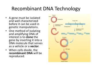
Cloning: To make identical copies DNA cloning involves separating a specific gene or DNA segment from a chromosome, attaching it to a DNA carrier molecule, and replicating this modified DNA, thousands or millions of times, through an increase in cell number and DNA copies per cell. The result is selective purification and amplification of a particular target gene or DNA segment from a complex mixture of DNA molecules. The methods used to accomplish these and related tasks are collectively referred to as recombinant DNA technology or genetic engineering.
Cloning vectors allow amplification of inserted DNA fragments • Developed from naturally occurring bacterial plasmids • Contain an origin of replication (ori ) • Contain numerous restriction sites • Contain genes that confer resistance to antibiotics, thus allowing selection of bacterial colonies carrying the plasmid • Introduced into competent bacterial cells by transformation
Different types of cloning vectors Plasmids: Circular DNA molecules which replicate separately from the host chromosome. Plasmids used for genomic and cDNA cloning. Bacterial host. Insert size range < 15kb. Bacteriophage-based Cosmids:Linear DNA molecules used for genomic and cDNA cloning. Bacterial host. Insert size range < 20kb. Bacterial Artificial Chromosomes (BACs): Circular DNA molecules used for cloning very long segments of genomic DNA. Bacterial host. Insert size range 100-300 kb. Yeast Artificial Chromosomes (YACs) : Specialized DNA molecules used for cloning very, very long segments of genomic DNA. Yeast host. Insert size range 100-2000kb.
Note : Multiple Cloning Sites (MCS) or a Polylinker Region Mammalian expression vector With the exception of budding yeast, plasmids are uncommon in eukaryotes. Thus, most eukaryotic vectors are based on DNA or RNA viral genomes. * * * * * * * Viral DNA sequences Bacterial sequences
Restriction endonucleases cut DNA molecules at defined positions A restriction enzymes binds to DNA at a specific sequence and make a double-stranded cut at or near that sequence.
Blunt and sticky ends Digestion of DNA with different restriction endonucleases 5’ and 3’ overhangs The same sticky ends produced by different enzymes
Amplification of a DNA Segment REQUIREMENTS: Oligonucleotide primers which flank the sequence of interest A DNA Template (a few ng) A thermal-stable DNA Polymerase (TAQ) dNTPs An automated thermocycler Long Product 5’ 5’ Long Product Polymerase Chain Reaction (PCR) DNA from a selected region of the chromosome or genome can to be amplified a billion-fold, effectively “purifying” it away from a complex mixture of DNA molecules. A repetitive three- step process : “Denature--Anneal--Elongate” (94-97oC) (42-55oC) (72oC)
Polymerase Chain Reaction (PCR) The Long Product (LP) acts as template for new synthesis SP LP LP Gives rise to Short Product (SP) whose 5’ and 3’ ends are both set by the primer annealing positions SP
Sequential rounds Polymerase Chain Reaction (PCR) SP SP In subsequent rounds, the Short Products accumulate in an exponential fashion
Following restriction digestion, the vector and insert are purified by agarose gel electrophoresis TET TET DNA ligation reaction is transformed into “competent cells” and then spread on selective agar plates
Log10 bp Cut EcoRI / Pvu II / Not I Vector [uncut] DNA Marker Cut EcoRI / Pvu II Cut EcoRI / Pvu II Clone 2 DNA Marker DNA Marker Distance Cut EcoRI / Pvu II Clone 2 Vector Insert EcoR I Pvu II Not I Analysis of Recombinant Clones: Restriction Enzyme Digestion 1.2% agarose gel cast In 1X TAE buffer DNA fragments stained with ethidium bromide and visualized by UV illumination. Vector Insert
Base PO4 Base PO4 H H H H OH H O O H H H H H H Analysis of Recombinant Clones: Thermal Cycle DNA Sequencing dNTP ddNTP
Genomic Library Construction using Bacteriophage l-based Vectors The l genome contains “optional” DNA * Cos site * * Genes are arranged into functional groups Insertion and Replacement Vectors Insert size range < 20 kb Cos sites incorporated into a plasmid = Cosmid
Genomic DNA Library Construction Analysis: Colony Hybridization Nytran or Nitrocellulose membrane Add an in vitro packaging mix
Restriction digestion Shotgun Sequencing Approaches A large segment of genomic DNA or a chromosome A whole genome Note: A genomic map is needed to provide a guide for sequencing by showing the positions of genes and other distinctive features. Closing a “sequencing gap”
Block II Lecture 1: Recombinant DNA Technology Part II. Experimental Problems and Approaches Assigning Genes to Chromosomal Locations Genetic Mapping RFLP and SSLP Analysis Physical Mapping Positional Cloning of a Target Gene cDNA synthesis and expression cloning Mapping Genes using ESTs Cloning Large Multigene Families by Degenerate PCR Cloning of a Target “Protein” and Physical Mapping
Genetic and Physical Mapping of a Gene to a Chromosome Genetic mapping enables physical mapping Genetic markers used for chromosomal mapping: Restriction site variation Repetitivesequences Genetic Linkage Analysis
Genetic Mapping Restriction Fragment Length Polymorphism (RFLPs) Useful molecular marker loci for chromosomal mapping and diagnosis of human disease genes This technique takes advantage of the ability of bacterial restriction enzymes to cut DNA at specific target sequences that exist randomly in the DNA of other organisms. Generally, the target sites are found at the same position in the DNA of different individuals within a population (i.e. the DNA of homologous chromosomes). Frequently, a specific site is missing because of some silent mutation. The mutation could be within a gene or a non coding intergenic region.
3kb 2kb 1kb 3 kb Homolog 1 3kb Homolog 2 3 kb If an individual is heterozygous for the presence (+) and absence (+/ -) of a restriction site, that locus can be used in mapping. The (+ / -) sites are detected by Southern blot analysis using a probe derived from that region. Southern blot analysis of this individual’s DNA would detect three fragments, 3, 2, and 1kb in length. 3 kb Homolog 1 Homolog 2 1 kb 2 kb Extent of probe Another individual might be homozygous for the long fragment and would show only a 3 kb band on a Southern blot. Southern blot analysis of this individual’s DNA would detect one fragment 3kb in length. Extent of probe Multiple forms of this region constitute an RFLP
2 kb 1 kb D d 3 kb 3kb 2kb 1kb 3kb Homolog 1 Homolog 2 In a cross of the two previous individuals, 50% of the progeny would show 3 fragments when probed, and the other 50% would show 1 fragment. This result follows Mendel’s Law of Equal Segregation, just as a gene would.
2 kb 1 kb D d 3 kb Homolog 1 Homolog 2 Hence, an RFLP can be mapped and treated like any other chromosomal site. Linkage of the heterozygous RFLP to a heterozygous gene with D coupled to the 1 plus 2 morph. Crossover between these sites would produce recombinant products (D-3, d-2-1). With this approach, the RFLP locus can be mapped relative to other molecular markers.
Restriction Fragment Length Polymorphism (RFLP) Analysis Evidence Suspect Victim “DNA Fingerprinting”used in modern forensics
Simple-Sequence Length Polymorphisms (SSLPs) D VNTRs : Variation in the Number of Tandem Repeats or “Mini-satellite” Molecular Markers d Probe binds repetitive sequences Restriction target sites are outside the repetitive array. The basic unit of the array is indicated by the arrows. The number of repeated units in a tandem array is variable. Individuals heterozygous for different numbers of tandem repeats can be detected, and the heterozygous site (s) used as a marker (s) for mapping. This VNTR locus will form two bands on a Southern blot: one long and one short. Similar to an RFLP locus, this heterozygous site can be used for genetic mapping. At present, VNTR analysis is rapidly performed using PCR.
Genetic profiling using Mini-Satellite VNTRs VNTRs located on the short arm of Chromosome 6 were amplified by PCR. The PCR Products were labeled with a blue or green fluorescent marker and resolved on a polyacrylamide gel. Each lane displays the genetic profile of a different individual. No two individuals will have the same genetic profile because each person had a different set of mini-satellite variants, which give rise to bands of different sizes after PCR. The red bands are DNA markers.
Positional cloning of a human target gene Contigs “Chromosomal Walking” technique used to identify single-disease genes in humans
cDNA Synthesis DNA molecules copied from an mRNA molecule by RT and therefore lack introns in genomic DNA Isolate mRNA from cell or tissue of interest Check integrity of RNA prep on HCHO gel Convert total pool of mRNA into cDNA using RT Clone cDNA into a DNA vector (e.g. l Zap) l to construct a cDNA expression library. Propagate and amplify cDNA library in a suitable host. Screen for cDNA of interest using DNA probe or antibodies that recognize the encoded protein.
ESTs are obtained by sequencing into the cDNA insert using a primer based on the vector sequence 5’ cDNA 3’ Gene Mapping using Expressed Sequence Tags (ESTs) EST DATABASE A collection of partial cDNA sequences, generally 200 to 400 bp in length, that was generated by sequencing vast numbers of cDNAs isolated from human cells and important model organisms such as mouse, Drosophila, and Caenorhabditis elegans. Composed of relatively short portions (tags) of genomic DNA sequences that are expressed in the form of mRNA. The EST database is constantly updated as sequences from increasing number of cDNA clones are added.
Genetic code contains redundancies = Degenerate ATT-Ile TAT- Tyr TTA - Leu TTG- Leu CTT- Leu CTC- Leu CTG- Leu STOP Codons TAA TAG TGA 20 Different Naturally Occurring Amino Acids 64 CODONS : 61 encode amino acids
NH2 COOH tyr phe ile ser ser asn ser thr leu asn ala lys leu his leu thr Computer programs apply the triplet-based genetic code to translate the EST sequences into partial amino acid sequence. Three nucleotides (a codon) are read from a specific starting point. If a match is found, then the EST provides the unique DNA sequence of that portion of the cDNA. A single probe that is complementary to the portion of the EST can be used to screen a genomic DNA library; the probe could also be used to screen a cDNA library
Odorant Receptors and the Organization of the Olfactory System Cloning a Large Multi-Gene Family by Degenerate PCR
Experimental design based on three assumptions: Odorant receptors likely belong to a superfamily of receptors (i.e. seven transmembrane domain receptors) that transduce intracellular signals by coupling to GTP-binding proteins The large number of structurally distinct odorous molecules suggests that the odorant receptors themselves should exhibit significant diversity and are likely to be encoded by a multigene family. 3. Expression of odorant receptors should be restricted to the olfactory epithelium.
VII II 5’ primers (match Domain II sequences) 3’ primers (match Domain VII sequences) GOAL: To identify molecules in the olfactory epithelium that resemble members of the seven transmembrane domain superfamily. Step 1. Extract RNA from olfactory epithelium and prepare cDNA Step 2. cDNA is amplified by PCR using a series of degenerate oligonucleotide primers that anneal to conserved regions of members of the superfamily of G-coupled seven transmembrane domain receptor genes. Each of the five different 5’ primer was used in PCR Reactions with each of six different 3’ primers.
Step 3. The amplification products of each PCR reaction were analyzed by agarose gel electrophoresis Step 4. PCR products within the size range expected for this family of receptor (600-1300 bp) were selected for further amplification with the appropriate primer pair to isolate individual bands. Each of the semi-purified PCR products was digested with the restriction enzyme Hinfl and analyzed by gel electrophoresis. (22 of the 64 PCR products isolated) PCR 13 yields a very large number of restriction fragments whose molecular weight sums to a value 5- to 10-fold greater than the original PCR product (13 different species of DNA)
Step 5. PCR 13 DNA was cloned into the plasmid vector Bluescript and 5 clones analyzed by DNA sequencing Each clone exhibited a different DNA sequence, BUT each encoded a protein that displayed conserved features of the superfamily of seven transmembrane receptor proteins. The proteins encoded by all 5 clones shared distinctive sequence motifs not found in other superfamily members , indicating they were all members of a NEW family of receptors Step 6. Obtain full-length cDNA clones by screening cDNA libraries prepared from olfactory epithelium RNA or RNA from enriched populations of olfactory sensory neurons Primary screen used a mixture of PCR 13 DNA as the probe (20 positives) Secondary screen used the original pair of primers used to amplify PCR 13 DNA (A4/B6) Step 7. Confirm expression of isolated cDNAs is restricted to epithelium using Northern blot analysis RESULT: Identified 18 members of a novel, extremely large multi-gene family that encoded olfactory receptors and lead to future work that merited the 2004 Nobel Prize in Medicine.
COOH NH2 H2 N H2 N H2 N COOH COOH COOH H2 N COOH COOH H2 N Cloning of a “target protein X” Digest with protease Step 1 Separate peptides • Protein X • Encoded by a pathogen • Gene locus unassigned Step 2 Step 3 Automated peptide sequencing
Design a “degenerate” probe based on partial protein sequence Mixture of 96 oligonucleotides that encode a portion of the peptide Degenerate PCR Note: If a Protein X EST database existed, you could design a single probe that was based on partial protein sequences • Once DNA sequence of the target gene is available, you could: • Map the entire gene and its location within the pathogen genome • Clone and sequence the transcript(s) encoded by the Protein X gene • Define the Protein X gene structure • Construct expression plasmids for functional studies of Protein X in cells • Mutagenize the Protein X cDNA using PCR-based site-directed mutagenesis and perform structure-function analysis • Produce recombinant protein X for vaccine development studies
REFERENCE MATERIALS FOR BLOCK 2/ LECTURE 1/ DNA Manipulations Lehninger Principles of Biochemistry, 3rd edition, Chapter 29 An Introduction to Genetic Analysis , 7th edition, Chapters 6, 7, 12, and 13 (http://WWW.WHFREEMAN.COM/BIOLOGY) FYI Lab Math: A handbook of Measurements, Calculations, and Other Quantitative Skills for Use at the Bench. D.S. Adams (2003) CSH Laboratory Press.