BCL2L10 & ER-GFP Expression in Mouse Muscle Fibers
Investigating BCL2L10 & ER-GFP expression patterns in mouse skeletal muscle fibers through confocal imaging. Includes data on BCL2L10 overexpression causing mitochondrial depolarization. Methods for transfection and fiber isolation provided.

BCL2L10 & ER-GFP Expression in Mouse Muscle Fibers
E N D
Presentation Transcript
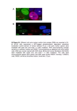
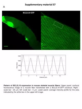
Supplementarymaterial S7 A BCL2L10-GFP • Pattern of BCL2L10 expression in mouse skeletal muscle fibers: Upper panel: confocal fluorescence image of a muscle fiber transfected with a BCL2L10-GFP construct. Right : scale bar : 20 m; left: scale bar : 5 m. Lower panel: average intensity profile for the area indicated by the white box in the upper left image.
Supplementarymaterial S7 B ER-GFP • Pattern of expression of GFP targeted to the sarcoplasmic reticulum (ER-GFP) in mouse skeletal muscle fibers: Upper panel: confocal fluorescence image of a muscle fiber transfected with an ER-GFP construct. Right : scale bar : 20 m; left: scale bar : 5 m. Lower panel: average intensity profile for the area indicated by the white box in the upper image.
Supplementarymaterial S7 C BCL2L10-GFP TMRM Merge • Overexpression of BCL2L10 in mouse skeletal muscle fibers causes mitochondrial membrane depolarization: Confocal fluorescence images of a skeletal muscle fiber transfected with BCL2L10-GFP and loaded with TMRM. The upper left image shows the BCL2L10-GFP fluorescence. The upper right image shows the TMRM fluorescence. The bottom image shows the two merged images. Scale bar: 20 μm.
Material and Methods for Supplementary material S7: • Ethical approval • Experiments were performed on 5–8-week-old male OF1 mice (Charles River Laboratories, L'Arbresle, France). All experiments and procedures were in accordance with the guidelines of the French Ministry of Agriculture (87/848) and of the European Community (86/609/EEC). They were approved by the local animal ethic committee of Rhone-Alpes, approval numbers 692660602, 0292. • Transfection of BCL2L10 in adult mice FDB muscles by electroporation • In vivo transfection of BCL2L10-GFP was performed within the flexor digitorumbrevis (FDB) of the animals, according to Weiss et al, 2008. In brief, mice were anaesthetized by intraperitoneal injection of a mixture of ketamine (Merial, Lyon, France) and xylazine (Bayer, Kiel, Germany) dissolved in sterile saline (80 and 16 mg kg−1, respectively). A 50 μl aliquot of a 10 μg μl−1 solution of BCL2L10-GFP (in sterile saline) was injected subcutaneously in the ventral side of the hindlimb paws through a 29-gauge needle (Terumo, Leuven, Belgium). The paw was then placed between two flat platinum electrodes. The standard protocol consisted of eight pulses of 200 V cm−1 amplitude of 20 ms duration at 1 Hz (ECM 830 Electro Square Porator, BTX). Experimental observations and measurements were carried out 5 days later. • Isolation of single FDB fibers • Single fibers were isolated from the mouse FDB muscles using a previously described procedure (Jacquemond, 1997) In brief, mice were killed by cervical dislocation. Muscle were removed and placed in a Tyrode solution containing 0.2% collagenase (Sigma, type 1) for 50 min at 37°C. After this treatment, muscles were kept at 4 C in Tyrode and used within 10 hours. Single fibers were obtained by triturating the muscles within the experimental chamber. For observations, fibers were bathed in Tyrode. • Solution and dye • Tyrode solution contained (in mM): 136 NaCl, 5 KCl, 2.6 CaCl2, 1 MgCl2, 10 Hepes, 10 Glucose, 5 Pyruvate. • Loading of the mitochondrial potential probe was carried out at room temperature in Tyrode. Cells were loaded with 10 nMTetramethylRhodamine Methyl Ester (TMRM, Molecular Probes, Eugene, OR, USA) for 10 min then washed with Tyrode containing 2.5 nM TMRM to avoid loss of the dye. • Confocal imaging and image processing • Experiments were performed at room temperature. Fibers were visualized using a Zeiss LSM 5 Exciter laser scanning confocal microscope (Zeiss, Jena, Germany) equipped with a ×63 oil immersion objective ×1.4 numerical aperture. For detection of BCL2L10-GFP, the excitation was provided by an argon laser (488 nm) and fluorescence was collected between 505 and 545 nm. For detection of TMRM, the excitation was from the 543 nm line of a HeNe laser and fluorescence was collected above 560 nm. Frame size was 512×512 pixels, with a pixel depth of 12 bit. Pinhole was set to 1 airy unit. • Data processing • Image and data processing were performed using Image/J (NIH, USA). • Jacquemond V. Indo-1 fluorescence signals elicited by membrane depolarization in enzymatically isolated mouse skeletal muscle fibers. Biophys J 1997,73:920–928. • Weiss N, Couchoux H, Legrand C, Berthier C, Allard B & Jacquemond V. Expression of the muscular dystrophy-associated caveolin-3(P104L) mutant in adult mouse skeletal muscle specifically alters the Ca2+ channel function of the dihydropyridine receptor. Pflugers Arch 2008, 457:361–375.