Analysis of Cell Cycle Markers for Double-Strand Break Resection in G1 and G2 Phase
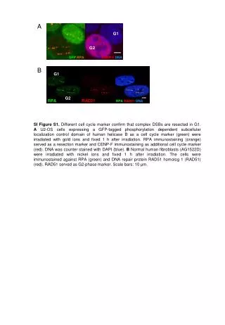
This study investigates the presence of complex double-strand breaks (DSBs) during the G1 phase of the cell cycle using AU2-OS cells expressing a GFP-tagged helicase B. Cells were irradiated and subjected to immunostaining for RPA as a resection marker and CENP-F as an additional cell cycle marker. Furthermore, normal human fibroblasts (AG1522D) were analyzed for RPA and RAD51 expression following nickel ion irradiation, with RAD51 serving as a G2 phase marker. Results showcase effective localization of markers, with a focus on understanding DSB resection.

Analysis of Cell Cycle Markers for Double-Strand Break Resection in G1 and G2 Phase
E N D
Presentation Transcript
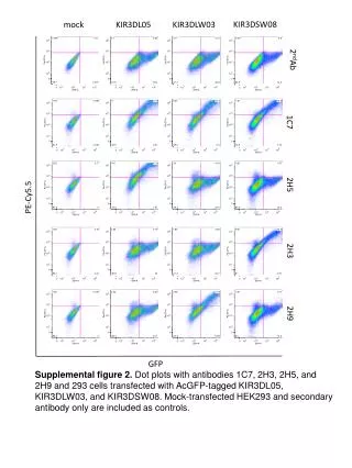
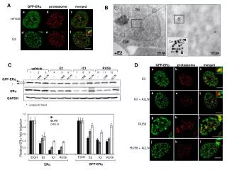
G1 G2 GFP RPA CENP-FDNA G1 c G2 RAD51 RPA RPA RAD51DNA A B • SI Figure S1. Different cell cycle marker confirm that complex DSBs are resected in G1. AU2-OS cells expressing a GFP-tagged phosphorylation dependent subcellular localization control domain of human helicase B as a cell cycle marker (green) were irradiated with gold ions and fixed 1 h after irradiation. RPA immunostaining (orange) served as a resection marker and CENP-F immunostaining as additional cell cycle marker (red). DNA was counter stained with DAPI (blue). B Normal human fibroblasts (AG1522D) were irradiated with nickel ions and fixed 1 h after irradiation. The cells were immunostained against RPA (green) and DNA repair protein RAD51 homolog 1 (RAD51) (red). RAD51 served as G2-phase marker. Scale bars: 10 μm.