Download
1 / 36
370 likes | 486 Vues
Explore the intricate relationship between cytoskeleton filaments and cancer progression, including how these filaments play a crucial role in cancer genesis, cell metastasis, and treatment approaches. Understand the molecular basis of cancer and the mechanisms involved in cancerous cell growth. Discover how cytoskeleton dynamics influence cancer development and potential therapeutic interventions.

E N D
ROLE OF CYTOSKELETON FILAMENTSIN CANCER Jigar S. Mehta M.Sc. Biochemistry (Semester-1) Saurashtra University Rajkot
Content • INTRODUCTION • Brief idea about 1) CYTOSKELETON - cytoskeleton filaments - functions of cytoskeleton. 2) CANCER - causing of cancer - Characteristics of Cancer cells - Molecular bases of Cancer - treatments of Cancer • PRINCIPLE ROLE OF CYTOSKELETON FILAMENTS IN CANCER I. Cancer genesis (APC beta-catenin pathway) II. Cancer cell metastasis III. Treatment of Cancer. • SUMMARY • RESEARCH PAPERS AND REFERENCES
Introduction • What do we mean by the sentence, -” Role of cytoskeleton filaments in Cancer” ? • Positive role OR Negetive role ? & • How it plays the role ?
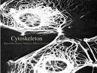
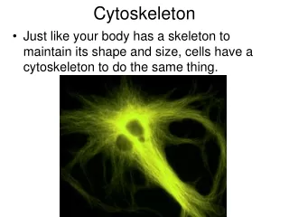
Cytoskeleton • Cytoskeleton : The ability of eucaryotic cells to organize the manycomponents in their interior, to adopt a variety of shapes, and to carry out coordinated movements depends on the cytoskeleton- an intricate network of protein filaments that extends throughout the cytoplasm. • This network of filaments helps to support the large volume of cytoplasm in a eucaryotic cell, a function that is particularly important in animal cells, which have no cell walls. Unlike our own bony skeleton, however, the cytoskeleton is a highly dynamic structure that is continuously reorganized as a cell changes shape, divides, and responds to its environment. • The cytoskeleton is not only the “bones” if a cell but its “muscle” too, and it is directly responsible for large-scale movements such as the crawling of cells along a surface, contraction of muscle cells, and the changes in cell shape that take place as an embryo develops.
Cytoskeletonfilaments • Cytoskeleton consist of :- a. microfilaments. (actin and myosin). b. intermediate filaments. (keratin-epithelial cell, vimentin-fibroblast,desmin-muscles, neurofilament-glial cells) c. microtubules. (tubulin).
cancer • Derivation of the term “Cancer” : Cancer is the common term for all malignant tumors. • Although the ancient origins of this term are somewhat uncertain, it probably derives from the Latin for crab, cancer—presumably because a cancer “ adheres to any part that it seizes upon in an obstinate manner like the crab.” • Cancerous condition : The process of celldivision in the body is a specifically controlled process. This helps in maintaining a balance between the cells dying and the cells reformed, in the tissues and organs of the body. Sometimes, in some part of the body, this process becomes uncontrolled due to some reason. This results in the development of a large cell mass which is called-tumour(tumor). Tumours can be of two types – Benign tumour is surrounded by connective tissue and it is localized. The tumuor does not grow indefinitely. A malignant tumour is not localized at one place. The cells of such a tumour become separated and spread through blood or lymph to other parts of the body and develop new malignant tumours in those organs. This condition in the body is known as cancerous condition
Characteristicsof cancerous cells • They undergo uncontrolled cell division. • The need of extracellular growth stimulents decreases.= self sufficiency in growth signals. • Defects in DNA repair. • These cells can migrate to new locations, establish themselves there and continue to divide uncontrollably and develop new malignant tumours. i.e. ability to invade and metastsize. • They show great difference in their cellular surfaces, cyto plasmic constitution and genetic constitution.
Causes of cancer • Cancer is caused by physical, chemical and biological factors. These factors called – carcinogens. • There are cancer causing genes called oncogenes in cells. • Under some specific conditions, they become active and form cancerous tumour. • Many polluants, ultraviolet rays and other radiations as well as several viruses are considered responsible for causing cancer. • “One of the important factor is mutation due to mutagens”.
MOLECULAR BASIS OF CANCER ACQ,(ENV) DNA damaging agents: Chemicals, radiation,viruses NORMAL CELL SUCCESSFUL DNA REPAIR DNA damage INHERITED MUTATIONS IN -GENES AFFECTING DNA REPAIR -GENES AFFECTING APOPSTOSIS FAILURE OF DNA REPAIR MUTATIONS IN GENOME OF SOMATIC CELLS ACTIVATION OF GROWTH PROMOTING ONCOGENES INACTIVATION OF TUMOR SUPPRESSOR GENES ALTERATIONS IN GENES THAT REGULATE APOPSTOSIS UNREGULATED CELL PROLIFERATION DECREASEDAPOSTOSIS CLONAL EXPANSIION ANGIOGENESIS ADDITIONAL MUTATIONS ESCAPE FROM IMMUMNITY TUMOR PROGRESSION INVASION AND METASTASIS MALIGNANT NEOPLASM
TREATMENT OF CANCER • SURGERY(removal of cancerous organ ) • RADIATION THERAPY ( arrest of cancer cell proliferation through cobalt rays) • CHEMOTHERAPY( anticancer drugs) • IMMUNOTHERAPY (hybridoma technique)
PRINCIPLE ROLE OF CYTOSKELETON IN CANCER • CANCER GENESIS or CARCINOGENESIS (APC beta catenin pathway) • CANCER CELL MOTILITY/METASTASIS • CANCER TREATMENT (chemotheraphy)
CANCER GENESIS / CARCINOGENESIS(the APC beta catenin pathway)
CARCINOGENESIS In our body cell division is happening continuously- the cell division of somatic cell & cell proliferation. But it is not occurring in uncontrolled manner, but is pre programmed process. Thus the no. of cells in our body/organ is maintained, and if tumor is going to form then with the help of tumor suppressor gene it is prevented. There is a crucial role of DNA repair mechanism. If any defect occur, it can lead to mutations in cells, which can cause Tumor (undifferentiated cell mass and it leads to cancer). In cells, some mechanism are involved to prevent further proliferation of cells if cells, if they have achieved their programmed state. One such pathway is APC beta Catenin Pathway
APC beta Catenin Pathway Down regulation of growth promoting signals is another potential area in which products of the APC & NF 1 gene lies into this category. Germ line mutations at the APC & NF 1 are associated with benign tumor that are precursor of carcinoma that develop later. The molecular basis if APC action and the basis of it’s tumor suppresor activity have been learned by the study of homologous genes in the fruit fly Drosophila and the Amphibian Xenopus. APC is a component of the WNT signaling pathway, which has a major role in controlling cell fate adhesion, cell polarity during embryonic development. WNT signaling is also required for self renewal of hematopoietic stem cells. WNT signals through a family of cell surface receptors called frizzeled(FRZ) stimulates several pathways. Central one involving beta Catenin And APC pathway. `
Normal(Somatic) Cells WNT WNT RECEPTOR E- CADHERIN beta catenin beta catenin Destruction complex beta catenin APC APC SIGNALS TFC beta catenin CELL WITHOUT WNT SIGNALS CELL CONT. WNT SIGNALLING No PROLIFERATION PROLIFERATION
CANCERCELLS (MUTATED) beta catenin APC NO EFFECT OF EXTERNAL STIMULI ON CANCER CELLS beta catenin PROLIFERATION
CANCER CELL MOTILITY “Cancer cell metastasis is a muti stage process involving invasion into surrounding tissue, intravasation, tansit in the blood or lymph, extravasation and growth at a new site. Many of these steps require cell motility, which is driven by cycles of actin polymerization, cell adhesion and acto-myosin contraction.” • Due to this motility, the invade other tissues, organs and spread cancer. • Normally cells have their motility according to the external stimuli through receptor molecules/chemicals and stop their motility after the stimuli is over. • In Cancer cells, as we are knowing, there is no external stimuli, so they continue and discontinue their motility according to their wish. • The cytoskeleton filament actin plays role in motility of cells through polymerization and depolymerization. • Above 5% of the total protein in the typical anima cell is Actin; about half of this is assembled into acctin filaments, while the other f\half is free as actin monomers in cytosol. The concentration of monomer is therefore high-much higher than the concentration required for purified action monomers to polymerize in vitro.
“What then keeps the actin monomers in cell from polymerizing totally into filaments?” The answer is that cells contain small proteins, such as thymosin and prolifin, that bind to actin monomers in the cytosol, preventing them from adding to the ends of actin filaments. By keeping actin monomers in reserve untill they are required, thymosin in particular plays a crucial role in regulating actin polymerization in cells. • In cancer cells, this proteins are being mutated, so their functions are lost/altered. • This leads to continuous polymerization and de polymerization of actin filamnets and thus self perform continuous crawling/uncontrolled motility, and change their location leading to METASTASIS which is known as Cancerous Cell Metastasis.
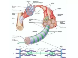
Cytoskeleton plays a very critical role in the chemotherapic method for the treatment of cancer . • The filaments microtubules and actin take part in the drug action • Drugs acting on this filaments are: • anticancer alkaloids and taxols,colchicine,vinca alkaloids(vinblastine and vincristine) • Most widely used are TAXOL (PACLITAXEL) & COLCHICINE
MECHANISM OF ACTION Paclitaxel(TAXOLS) interferes with the normal function of microtubule breakdown. Whereas drugs like colchicines cause the depolymerization of microtubules, paclitaxel arrests their function by having the opposite effect; it hyper-stabilizes their structure. This destroys the cell's ability to use its cytoskeleton in a flexible manner. Specifically, paclitaxel binds to the β subunit of tubulin. Tubulin is the "building block" of microtubules, and the binding of paclitaxel locks these building blocks in place. The resulting microtubule/paclitaxel complex does not have the ability to disassemble. This adversely affects cell function because the shortening and lengthening of microtubules (termed dynamic instability) is necessary for their function as a mechanism to transport other cellular components. For example, during mitosis, microtubules position the chromosomes during their replication and subsequent separation into the two daughter-cell nuclei. Further research has indicated that paclitaxel induces programmed cell death (apoptosis) in cancer cells by binding to an apoptosis stopping protein called Bcl-2 (B-cell leukemia 2) and thus arresting its function.
In addition to stabilizing microtubules paclitaxel may act as a molecular mop by sequestering free tubulin effectively depleting the cells supply of tubulin monomers and/or dimers. This activity may trigger the aforementioned apoptosis. One common characteristic of most cancer cells is their rapid rate of cell division. In order to accommodate this, the cytoskeleton of a cell undergoes extensive restructuring. Paclitaxel is an effective treatment for aggressive cancers because it adversely affects the process of cell division by preventing this restructuring. Cancer cells are also destroyed by the aforementioned anti-Bcl-2 mechanism. Other cells are also affected adversely, but since cancer cells divide much faster than non-cancerous cells, they are far more susceptible to paclitaxel.
SUMMARY Cancer is a disease in which many of the characteristics of normal cell behavior are lost or perturbed. Uncontrolled cell proliferation and inappropriate cell survival are common features of all cancers, but in addition defects in cellular morphogenesis that lead to tissue disruption, the acquisition of inappropriate migratory and invasive characteristics and the generation of genomic instability through defects in mitosis also play important role in the progression of the disease. Actin and tubulin form highly versatile, dynamic polymers that can organize cytoplasmic organelles and intracellular compartments define cell polarity and generate bpth pushing and contractile forces. Therefore it is not surprisind that they are key players in many processes in cell biology. In the cell cycle, these two cytoskeletal structures drive chromosomal seperation and cell division. During morphogenesis, they determine cell shape and polarity, and promote stable cell-cell and cell-matrix adhesion through their interactions with cadherins and integrins, respectively.
Finally, during cell migration they generate protrusive forces at the front and retraction forces at the rear. These are all aspects of cell behavior that often go awry in cancer.
British Journal of Cancer (2002) 86, 1316–1321. Modulation of colony stimulating factor release and apoptosis in human colon cancer cells by anticancer drugs S Calatayud, T D Warner and J A Mitchell Abstract: Modulation of the immune response against tumour cells is emerging as a valuable approach for cancer treatment. Some experimental studies have shown that secretion of colony stimulating factors by cancer cells reduces their tumorigenicity and increases their immunogenicity probably by promoting the cytolitic and antigen presenting activities of leukocytes. We have observed that human colon cancer cells (HT-29) are able to secrete granulocyte-macrophage-colony stimulating factor, granulocyte-colony stimulating factor and macrophage-colony stimulating factor when stimulated with cytokines (IL-1and TNF-). In this study we assessed, for the first time, the effects of several anticancer drugs on colony stimulating factor release or apoptosis in HT-29 cells. Cytokine-induced release of granulocyte-macrophage-colony stimulating factor, granulocyte-colony stimulating factor and macrophage-colony stimulating factor was significantly increased by cisplatin and 6-mercaptopurine. Taxol only increased macrophage-colony stimulating factor release while reduced that of granulocyte-colony stimulating factor. No changes in colony stimulating factor secretion were observed after treatment with methotrexate. Only cisplatin and taxol induced apoptosis in these cells. Secretion of colony stimulating factors by colon cancer cells may contribute to the immune host response against them. Anticancer drugs such as cisplatin and 6-mercaptopurine increase colony stimulating factor secretion by cytokine stimulated cancer cells probably through mechanisms different to those leading to cell apoptosis, an effect that may contribute to their anti-neoplasic action.
Journal of Investigative Dermatology (1975) 65, 127–142. DESMOSOMES, FILAMENTS, AND KERATOHYALINE GRANULES: THEIR ROLE IN THE STABILIZATION AND KERATINIZATION OF THE EPIDERMIS A Gedeon Matoltsy Abstract Components of desmosomes, filaments, and keratohyaline granules were studied by electron microscope and biochemical methods to clarify their role in the stabilization and keratinization of the epidermis. Isolated desmosomes are composed of 76% protein, 17% carbohydrate, and 10% lipid. The bulk of protein consists of a "spectrin"-like fibrous protein, presumably present in the plaque, and of glycoproteins in the desmosomal interspace. The main component of filaments, prekeratin, is a low-sulfur -protein composed of a pair of three-chain subunits with non--helical segments separated by 200 Å-long -helical regions. The major component of isolated keratohyaline granules, the amorphous particulate material, is formed by a high-sulfur protein with a single-type of polypeptide chain. polypeptide chains comparable to those found in prekeratin and keratohyaline granules were recovered from extracts of horny cells. Within the living part of the epidermis, filaments hypothetically form a cytoskeletal system which is anchored to desmosomes by a filamentous plaque protein. Glycoproteins are involved in the formation of strong junctions between the cells which enable the living part of the epidermis to respond as a whole to mechanical stress. The stratum corneum is stabilized by a similar system in a consolidated state which is less extensible. Horny cells are enveloped by a thickened membrane and the interfilamentous spaces are filled with various proteins, including the sulfur-rich amorphous protein found in keratohyaline granules.
Journal of the National Cancer Institute, Vol. 82, No. 15, 1247-1259, 1990Taxol: A Novel Investigational Antimicrotubule Agent Eric K. Rowinsky*, Lorranie A. Cazenave, Ross C. Donehower Abstract: Microtubules are among the most strategic subcellular targetsof anticancer chemotherapeutics. Despite this fact, new antimicrotubuleagents that possess unique mechanisms of cytotoxic action andhave broader antineoplastic spectra than the vinca alkaloidshave not been introduced over the last several decades—untilthe recent development of taxol. Unlike classical antimicrotubuleagents like colchicine and the vinca alkaloids, which inducedepolymerization of microtubules, taxol induces tubulin polymerizationand forms extremely stable and nonfunctional microtubules. Taxolhas demonstrated broad activity in preclinical screening studies,and antineoplastic activity has been observed in several classicallyrefractory tumors. These tumors include cisplatin-resistantovarian carcinoma in phase II trials and malignant melanomaand non-small cell lung carcinoma in phase I studies. Taxol'sstructural complexity has hampered the development of feasibleprocesses for synthesis, and its extreme scarcity has limitedthe use of a conventional, broad-scale screening approach forevaluation of clinical antitumor activity. However, taxol'sunique mechanism of action, its spectrum of preclinical antitumoractivity, and tumor responses in early clinical trials havegenerated renewed interest in pursuing its development.
REFERENCE BOOKS Cell and Molecular Biology, 8th Edition by E.D.P. De Robertis E.M.F. De Robertis Robbins and Cotran’s Pathologic Basis Of Disease, 7th Edition by Kumar, Abbas & Tausto Cell biology by Thomas Pollard & William Earnshaw Essential Cell Biology by Alberts, Bray, Johnson, Lewis, Raff, Roberts, Walter Textbook of Biology Part-2, Std.12th (GSHSEB)