BURNS
BURNS. PRESENTED BY SOUMYA SARA JOSEPH. DEMOGRAPHIC DATA. Name: Case No.3 MR No : 196537 Diagnosis: 15% Burn Age: 2 YRS Gender: Male. PHYSICAL ASSESSMENT. GENERAL ASSESSMENT :

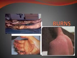
BURNS
E N D
Presentation Transcript
BURNS PRESENTEDBY SOUMYA SARA JOSEPH
DEMOGRAPHIC DATA Name: Case No.3 MR No : 196537 Diagnosis: 15% Burn Age: 2 YRS Gender: Male
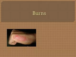
PHYSICAL ASSESSMENT GENERAL ASSESSMENT: Baby has fever, lethargic, dehydrated SKIN: Dry, No blisters, No edema, Right side upper chest , left elbow, right thigh 1st degree burn present, Dehydrated HEAD AND NECK: No Deformities Found
THORAX:Normally symmetrical in size • CARDIOVASCULAR: normal • GENITOURINARY: Diminished urine output
GASTROINTESTINAL : Abdomen is soft, not distended MUSCULO-SKELETAL : No deformities found NEUROLOGY : Growth and development is normal according to Erikson’s psycho social stage
PATIENT HISTORY Past Medical History No past history of any medical illness Present Medical History Now baby is admitted with complaints of 20% Burns
Topic Presentation BURNS
BURNS Tissue damage from excessive heat, electricity, radioactivity, or corrosive chemicals that destroys(denatures) proteins in the exposed cells is called a burn. Generally, the systemic effects of a burn are a greater threat to life than are the local effects.
FIRST-DEGREE – only epidermis (sunburn) SECOND-DEGREE BURN – destroys entire epidermis & part of dermis – fluid-filled blisters separate epidermis & dermis – epidermal derivatives are not damaged – heals without grafting in 3 to 4 weeks & may scar THIRD-DEGREE OR FULL-THICKNESS – destroy epidermis, dermis & epidermal derivatives – damaged area is numb due to loss of sensory nerves
STRUCTURE OF THE SKIN EPIDERMIS – The superficial portion of the skin – Composed of epithelial tissue. DERMIS – The deeper layer of the skin – Primarily composed of connective tissue
SUBCUTANEOUSOR HYPODERMIS – It consists of areolar and adipose tissue. – fat storage, an area for blood vessel passage, and an area of pressure sensing nerve endings.
EPIDERMIS Stratified squamous epithelium – Avascular (contains no blood vessels) – 4 types of cells – 5 distinct strata (layers) of cells
CELLS OF EPIDERMIS Four Principle Cells of the Epidermis • KERATINOCYTES – produce the protein keratin, which helps protect the skin and underlying tissue from heat, microbes, and chemicals, and lamellar granules, which release a waterproof sealant • MELANOCYTES – produce the pigment melanin which contributes to skin color and absorbs damaging ultraviolet (UV) light
LANGERHANS CELLS – derived from bone marrow – participate in immune response • MERKEL CELLS – contact a sensory structure called a tactile (Merkel) disc and function in the sensation of touch
LAYERS OF THE EPIDERMIS From deepest to most superficial the layers of the epidermis are – STRATUM BASALE (stratum germinativum) – STRATUM SPINOSUM – STRATUM GRANULOSUM – STRATUM LUCIDUM (only in palms and soles) – STRATUM CORNEUM
DERMIS • Connective tissue layer composed of collagen & elastic fibers, fibroblasts, macrophages & fat cells • Contains hair follicles, glands, nerves & blood vessels • Two major regions of dermis – papillary region – reticular region
ETIOLOGY • (from steam, hot bath water, tipped-over coffee cups, hot foods, cooking fluids, etc.) • Contact with flames or hot objects (from the stove, fireplace, curling iron, etc.) • chemical burns (from swallowing things, like drain cleaner or watch batteries, or spilling chemicals, such as bleach, onto the skin) • Electrical burns (from biting on electrical cords or sticking fingers or objects in electrical outlets, etc.) • overexposure to the sun
PATHOPHYSIOLOGY When large part of the body burnt this will effect most systems of the body.
SKELETAL SYSTEM Burn area is too large Destruction of red blood cells Blood transfusion Bone marrow replacement
MUSCULAR SYSTEM In case of burn Body becomes hyper metabolic
CARDIO-VASCULAR SYSTEM Burning of the skin Increase in capillary permeability Increase in blood vasculaturiture Decrease of Blood pressure Decreases blood flow and oxygenation to tissue Edema, shock and eventually death
NERVOUS SYSTEM Partial thickness burn Full thickness burn Only pain Nerve cell destruction no feeling Abnormal levels of circulating potassiumions Such as Cellular destruction, outward flow of K+ fluid in burn HYPERKALEMIA HYPOKALEMIA Transmission of messages in the nervous system work faster orSlower than normal or not at all
RESPIRATORY SYSTEM Airway obstruction Gross edema of the throat Increased rate of respiratory rate Pulmonary edema
ENDOCRINE SYSTEM Increased secretion of adrenaline and nor adrenaline Increased body temperature Increased cell metabolism
LYMPHATIC SYSTEM Increased inflammation Grater strain on lymphatic system Pitting edema
IMMUNE SYSTEM Excessive strain on Burns area removing the first Lymphatic system line of infection defense Decreased response Increased infection
DIGESTIVE SYSTEM Potential hypovolemic state Decrease in blood availability in the intestinal lining and liver Increases nutrients requires to support metabolism Repair of damaged cells
URINARY SYSETEM Increased fluid loss Decreased urine output Potential for kidney damage Poor perfusion
SIGN AND SYMPTOMS BOOKED BASE 1ST DEGREE • Redness • Pain • Minor swelling 2nd DEGREE • Severe pain • Blister+ and if it breaks the area wet looking with a bright pink to cherry red color • Redness 3rd DEGREE • Little or No pain • Skin Dry, look waxy white, leathery, brown or charred
PATIENT MANIFESTED • Fever • Pain • Redness • Minor swelling
INTRVENTION • Promoting comfort • Promoting fluid intake and maintain nutrition • Promoting pain relief and psycho logic adjustment • Promoting family knowledge • Prevention of infection • Monitoring And Prevention Of Complication • Promoting home and community based care
BOOK BASED 1ST & 2nd DEGREE BURNS • Sterile dressings • Preventive Antiseptics • Preventive Antibiotics • Analgesics • Hydration- 3RD DEGREE BURNS • Sterile dressing • Preventive Antibiotics • Preventive Antiseptics • Hydration • Transfusion • Skin grafting
FLUID CALCULATION FOR BURN PATIENT ACCORDING TO PARKLAND FORMULA 4 X TBSA ( %OF BURN ) X WEIGHT ONE HALF OF THE REQUIREMENTS ARE GIVEN DURING THE FIRST 8 HRS THE REMINDER IS GIVEN OVER THE NEXT 16 HRS
LUND AND BROWDER CHAT IS USED TO DETERMINE TO EXTENT OF BURNS IN CHILDREN BECAUSE IT IS BASED ON AGE, THUS COMPENSATING FOR CHANGES BASED ON GROWTH
COMPLICATIONS ACUTE Infection Curling’s ulcer (Stress) { TBSA>20%} Acute gastric dilation { TBSA>20%} Renal failure Respiratory failure Post burn seizures
Hypertension • Central Nervous system dysfunction • Vascular Ischemia • Anxiety and complex pain • Anemia and malnutrition • Constipation and fecal impaction • Alteration in mood secondary to hospitalization