Inhibition of fH Binding to Meningococci by Polyanions
Study on the impact of sucrose octasulfate and heparin on fH binding to meningococci from different fHbp variant families. Results show differential fH and FHL-1 binding to Neisseria meningitidis and Neisseria gonorrhoeae PorB.1A strains.

Inhibition of fH Binding to Meningococci by Polyanions
E N D
Presentation Transcript
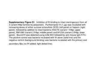
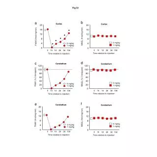
Supplementary Figure S1. Inhibition of fH binding to intact meningococci from all 3 variant fHbp families by polyanions. Purified factor H (1 µg) was incubated with increasing doses of either sucrose octasulfate (SOS) (left panel) or heparin (right panel), followed by addition to intact bacteria (H44/76 (variant 1 fHbp; upper panel), RM1090 (variant 2 fHbp; middle panel) and M1239 (variant 3 fHbp; lower panel). Bound fH was detected using mAb 90X followed by anti-mouse IgG-FITC. The positive control was bacteria incubated with fH alone (solid line) and the negative control (background binding) was bacteria incubated with the primary and secondary Abs (no fH added; light dotted line).
Supplementary Fig. S1 SOS Heparin H44/76 (variant 1) RM1090 (variant 2) M1239 (variant 3) control control fH fH fH+SOS 10mM fH+heparin 100 u/ml fH+SOS 40mM fH+heparin 400 u/ml
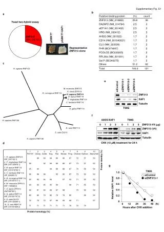
Supplementary Figure 2. Differential amounts of fH and FHL-1 binding to N. meningitidis and N. gonorrhoeae PorB.1A. N. meningitidis strain H44/76 (fHbp variant 1; left graph) and N. gonorrhoeae PorB.1A-expressing strain 15253 (right graph) were incubated with equimolar amounts (120 nM) of the full length purified fH and FHL-1. fH and FHL-1 bound to bacteria was detected using mAb 90X that is specific for fH SCR1 (and therefore also recognizes FHL-1), followed by anti-mouse IgG-FITC. Binding of the full length fH is shown by the shaded histograms, binding of FHL-1 by the solid lines and controls (no fH or FHL-1 added) by the broken lines. The x-axis represents fluorescence on a log10 scale and the y-axis the number of events on a linear scale.
Supplementary Fig. S2 N. meningitidis strain H4476 Variant 1 fHbp N. gonorrhoeae strain 15253 PorB.1A Isotype control Purified human factor H 18 µg/ml (~120 nM) Purified human FHL-1 5 mg/ml (~120 nM)

![[Fig. S1]](https://cdn3.slideserve.com/6448662/slide1-dt.jpg)