Trauma, Non Head, Non Spine
1.23k likes | 1.59k Vues
Trauma, Non Head, Non Spine. By CCM Fellows UBC. Case. -19 years old girl, otherwise healthy, was brought to ER at RCH by EHS after being hit by a freight train.

Trauma, Non Head, Non Spine
E N D
Presentation Transcript
Trauma, Non Head, Non Spine By CCM Fellows UBC
Case • -19 years old girl, otherwise healthy, was brought to ER at RCH by EHS after being hit by a freight train. • -Earlier, she was partying with her pals, got drunk, went through a fight with her bf, after which she decided to walk home alone! At the railway intersection, she was hit on her left side by the train, which slowed down coming near the station.
Upon EHS Arrival.. • -When EHS arrived within 2 minutes, she was conscious but drowsy, GCS E3 M5 (x4) V2, vomiting, with open wounds on her posterior scalp, and Lt knee. She was intubated at the seen and brought to ER.
In ER.. • A:ETT, C-collar. • B: AC, fiO2 0.5. ABG 7.3/50/19/88/ -6. • C: 110/50 (65), 110 SR, T:34C. • D: PERL 3mm bi, on 3:3 M:M + 100 mcg of fentanyl given by ER MD when patient was trying to wake up and bite on the ETT. • Trauma team are in.
O/E.. • HEENT: 3-cm Laceration wound grade I over the occiput. • Heart: Normal S1+S2. • Chest: paradoxical movement of Lt 3-7 ribs chest wall, with multiple bruises on the Lt side, decreased B/S on the Lt side. • Abdomen: multiple bruises on the Lt side, with mildly distended abdomen. DRE: clear. • F/C hematuria, 70cc/hr (BWt 90kg). • Ext: bruises over Lt shoulder posteriorly. Intact/symmetrical upper pulses/BP. Lt thigh swelling, 15-cm Lt knee grade IIIb (at least) laceration with exposed fractured bone, Lt PT and DP weaker than Rt
FAST.. • No tamponade, “good” LVEF, coarse spleen, evidence of fluid in the hepatorenal space and Lt perinephric area.
Labs.. • Hb 60, Plt 80, INR 1.5, PTT 40, Fib 0.9, WBC 16, • Cr 80, BUN 7, lytes N, LFE N. LA 4.3, Trop <0.04, • U/A RBC 20-50, WBC 5-10. • ECG: Sinus tachycardia.
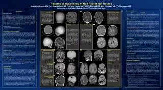
Imaging.. • CT head: N. C-spine: N. • Chest: Lt hemopneumothorax, fractured Lt scapula and ribs 3-7, Lt lung contusion. • T-L-S spine: intact. • Abdomen: Lt diaphragmatic rupture, spleen laceration grade 4, liver injury grade 1, Lt perinephric hematoma, evidence of hemoperitoneum. • Pelvis: Lt pubic ramus fracture. • Ext: Fracture Lt acetabular and femoral head capitus, displaced femoral shaft and intercondylar, patella and tibial plateau, with air tracking from the knee proximally, c/w open fracture. CTA preliminary report N. • -Lt chest tube was inserted, drained blood.
Question 1 • Discuss the initial fluid resuscitation in trauma patient, focusing of monitoring endpoints (including base deficit), coagulopathy (including hypothermia), and massive transfusion protocol (Marios)
Initial trauma fluid resuscitation Fluids Transfusion ratios Monitoring end-points Coagulopathy
Initial fluid resuscitation A controversial topic with nebulous answers Guidelines used to be that you gave 2 or more liters of crystalloid to any trauma patient you thought was in shock Goal was to rapidly restore circulating volume to maintain vital organ perfusion. There is evidence however that normalizing one’s blood pressure in the setting of an uncontrollable hemorrhage may worsen outcome. This has led to the concept of “permissive hypotension/hypotensive resuscitation”
Initial fluid resuscitation • Rationale behind permissive hypotension (animal models) • Increased blood pressure accelerates bleeding and dislodges soft early clots (which take 30 min to harden) • Dilution of RBC mass by crystalloid or colloid reduces oxygen delivery despite increasing cardiac output • Reduced hematocrit and clotting factor concentration inhibit clot formation • Resuscitative fluids themselves may have deleterious properties such as neutrophil activation (RL in hemorrhagic shock 1998)
Initial fluid resuscitation • Human evidence for permissive hypotension • Houston study (1994) • Thoraco-abdominal gunshot or stab wounds presenting with SBP < 90 mmHg • Patients either treated with “liberal” RL or delayed resuscitation until OR. • Patients in the early resuscitation group had a higher mortality and rate of post-op complications • Baltimore study (2002) • Penetrating and blunt trauma patients presenting to the Shock Trauma Center with SBP < 90 mmHg • Randomized to a fluid resuscitation strategy targeted to a lower than normal SBP (> 70 mmHg) or to conventional care (SBP > 100 mmHg) • Mortality was identical but fewer complications and a shorter duration of hemorrhage were seen in the low-pressure group Bickell WH, Wall MJ Jr, Pepe PE, et al. Immediate versus delayed fluid resuscitation for hypotensive patients with penetrating torso injuries. N Engl J Med 1994; 331:1105–1109. Dutton RP, Mackenzie CF, Scalea T. Hypotensive resuscitation during active hemorrhage: impact on in-hospital mortality. J Trauma 2002; (52):1141– 1146.
Initial fluid resuscitation • Who’s practicing permissive hypotension? • Current military policy is to resuscitate to a palpable radial pulse or an SBP of no more than 90 mmHg. • This concept has now been adopted widely, and is reflected in the latest EAST guidelines. Dawes R, Thomas GO. Battlefield resuscitation. Curr Opin Crit Care. 2009Dec;15(6):527-35.
Initial fluid resuscitation • Prehospital resuscitation (EAST guidelines 2009) • Obtaining IV access in the field has not been shown to be beneficial, and if anything has been shown to prolong transport time • Obtaining IV access en-route to trauma centre is recommended if wounds more than superficial. • IV should be saline-locked if no indication for fluid therapy is present. Cotton BA, Jerome R, Collier BR, et al. Eastern Association for the Surgery of Trauma Practice Parameter Workgroup for Prehospital Fluid Resuscitation. Guidelines for prehospital fluid resuscitation in the injured patient. J Trauma. 2009 Aug;67(2):389-402
Initial fluid resuscitation • Prehospital resuscitation (EAST guidelines 2009) • Indications for prehospital fluid administration (250 cc boluses) in both penetrating and blunt trauma: • Patient incoherent • Non-palpable radial pulse • Head injury with SBP < 90 • Repeat bolus if no response • Saline lock of pt responds
Initial fluid resuscitation • When may fluids be appropriate? • In traumatic brain injury, where hypotensive episodes have been associated with worse outcome • In severe hypotension where pressors would otherwise be needed, i.e. MAP < 40 - 50 mmHg • In hypotensive patients with controllable bleeding (extremity/superficial bleed)
If you’re going to bolus, bolus right. Right? Bolus with… • The data on what type of fluid to give is worse than on whether to give fluid. • EAST Guidelines states insufficient evidence to make recommendations. • Practice therefore depends on opinion: • RL is liked for its “buffering capacity” but can’t be mixed with blood due to the calcium • Plasmalyte doesn’t have calcium but has potassium which can exacerbate hyperkalemia secondary to tissue injury and massive transfusion • NS doesn’t have K+ (unless you ask for it), but is more likely to cause a NAGMA that can theoretically worsen coagulopathy • Hypertonic saline showed no benefit over isotonic crystalloids • Early availability of blood and FFP avoids the need for “filler fluids” Diez C, Varon AJ. Airway management and initial resuscitation of the trauma patient. Curr Opin Crit Care. 2009 Dec;15(6):542-7 Dawes R, Thomas GO. Battlefield resuscitation. Curr Opin Crit Care. 2009Dec;15(6):527-35.
Transfusion ratios • American and British military practice is to administer warmed FFP and PRBCs in a 1:1 ratio as soon as possible. • Others in military have recently suggested modifying this ratio by further adding platelets, resulting in a ratio of 1:1:1 PRBC:FFP:platelets • Evidence for benefit based on retrospective trials • Benefit can therefore be indicative of a survival bias rather than a true mortality benefit: • FFP and platelets take longer to receive than pRBCs. • Possibility that nonsurvivors did not die because they received a lower FFP : PRBC ratio, but that they received a lower ratio transfusion because they died. Dawes R, Thomas GO. Battlefield resuscitation. Curr Opin Crit Care. 2009Dec;15(6):527-35. Snyder CW, Weinberg JA, McGwin G Jr, et al. The relationship of blood product ratio to mortality: survival benefit or survival bias? J Trauma 2009;66:358–362;
Resuscitation endpoints • If uncontrolled hemorrhage: permissive hypotension, maintaining coherence, a palpable radial pulse, or an SBP > 90 mmHg in TBI • Resuscitation effectiveness can be assessed by standard measures, i.e. lactate clearance and correction of base deficit. • Base deficit: Blunt injury patients with transient field hypotension and a BD > 6 were found to be more than twice as likely to have repeat hypotension (crump). Bilello JF, Davis JW, Lemaster D, et al. Prehospital Hypotension in Blunt Trauma: Identifying the "Crump Factor". J Trauma. 2009 Dec 4 Tisherman SA, et al. Clinical practiceguideline: endpoints of resuscitation. J Trauma. 2004 Oct;57(4):898-912.
Hemorrhagic coagulopathy Part of the “lethal triad”
Hemorrhagic coagulopathy Impaired hemostasis is often caused by dilution and consumption of clotting factors and hyperfibrinolysis. However despite replacing FFP, platelets, and cryoprecipitate, patients may remain coagulopathic. Optimal coagulation requires specific preconditions concerning acid-base balance, calcium, hematocrit, and temperature. If these preconditions are not fulfilled, coagulation may remain abnormal despite replacement of products. Lier H, Krep H, Schroeder S, Stuber F. Preconditions of hemostasis in trauma: a review. The influence of acidosis, hypocalcemia, anemia, and hypothermia onfunctional hemostasis in trauma. J Trauma. 2008 Oct;65(4):951-60.
Hemorrhagic coagulopathy • Acidosis: • A notable impairment in hemostasis arises at pH <= 7.1 or a base deficit of 12.5 or more • Aggressive resuscitation in OR to reverse acidosis • Some centres give THAM to raise pH to 7.2 or higher (no outcome data) • Hypocalcemia: • Coagulation defects can be attributed to hypocalcemia if the Cai++ is < 0.6 – 0.7 mmol/L • Adverse cardiac effects commence at levels at or below 0.8 – 0.9 mmol/L • Combining these benefits, ionized calcium should be kept above 0.9 mmol/L Lier H, Krep H, Schroeder S, Stuber F. Preconditions of hemostasis in trauma: a review. The influence of acidosis, hypocalcemia, anemia, and hypothermia onfunctional hemostasis in trauma. J Trauma. 2008 Oct;65(4):951-60.
Hemorrhagic coagulopathy • Anemia: • Causes demargination of platelets and decreased adhesion to endothelial damage (decreases fivefold from HCT of 40% to 10%) • Aim is to keep HCT greater or equal to 30% • Hypothermia • High risk of persistent coagulopathy at temperatures under 35 deg C • At temperatures below 33 deg C, hypothermia produces a coagulopathy that is equivalent to 50% of normal activity at normothermia • Should therefore aggressively aim for a Temp > 34 or even 36 degrees Celsius Lier H, Krep H, Schroeder S, Stuber F. Preconditions of hemostasis in trauma: a review. The influence of acidosis, hypocalcemia, anemia, and hypothermia onfunctional hemostasis in trauma. J Trauma. 2008 Oct;65(4):951-60. Dawes R, Thomas GO. Battlefield resuscitation. Curr Opin Crit Care. 2009Dec;15(6):527-35.
Hemorrhagic coagulopathy • Other measures: • Hypofibrinogenemia keep fibrinogen > 1 g/L • Platelets keep above 100 x 109 • Tranexamic acid • At 15 mg/kg, found to reduce blood loss in elective surgical patients by inhibiting fibrinolysis. • Results of CRASH II trial are pending (20 000 patients randomized to 1 g of tranexamic acid followed by 1 g infused over 8 h). • rFVIIa • Some evidence that it reduced transfusion requirement in blunt injury but not in penetrating injury. • Often used in salvageable patients with continuing haemorrhage that has failed surgical and nonsurgical methods Mannucci PM, Levi M. Prevention and treatment of major blood loss. N Engl JMed. 2007 May 31;356(22):2301-11. Dawes R, Thomas GO. Battlefield resuscitation. Curr Opin Crit Care. 2009Dec;15(6):527-35.
-After transfusing 4u of PRBCs, 4u FFP, 10u Plt, 4u cryoppt, BP dropped to 80/45, O2 sat 80%, decreased B/S on Lt, with Lt CT suddenly draining >1500 ml of blood.
Question 2 • What are the indications and contraindications for ED thoracotomy? How to manage lung contusion and flail chest? What are the complications of lung contusion? (Erik)
Emergency Department Thoracotomy • Indications • Contraindications • Absolute • Relative
Objectives of EDT Release of pericardial tamponade. Control of intrathoracic vascular or cardiac bleeding. Evacuate obstructive air embolism or control source of broncho-pleural/vascular fistula. Perform open cardiac massage. Temporarily occlude the descending thoracic aorta.
Indications for EDT Penetrating chest injury in extremis, or loss of vital signs, within 10 minutes of ED arrival. Limited evidence to support in blunt or mutli-trauma patients, especially if arrive in ED with VSA. Known tamponade, air embolism. Consider in major abdominal vascular injury (blunt or penetrating) in extremis or witnessed loss of vital signs. Consider in unresponsive hypotension (SBP < 60mmHg) or chest tube > 1500cc*.
Contraindications Severe TBI VSA in penetrating injury > 10-15 minutes prior to ED arrival. VSA in blunt injury 0-5 minutes prior to ED arrival.
Pulmonary Contusion & Flail Chest Both PC and FC independently associated with morbidity. Pathophysiology Mortality usually resultant of other injuries sustained from the [blunt] trauma (e.g. CNS injury, shock).
Fluid Resuscitation in PC Animals models originally suggested that crystalloid resuscitation had greater impact versus colloid – but no outcomes were assessed. Similarly, observational data from Vietnam War suggested larger volume resuscitation was associated with poor outcomes. More recently, studies with better (though not great) methodology show no correlation with volume of resuscitation with worsening of PC. P/F ratio at the time of injury more prognostic.
Ventilation in PC/FC Again, animal models with inappropriate surrogate endpoints are misleading. Current level II evidence supports intubation and mechanical ventilation based on standard assessment of oxygenation/ventilation. Advantages of different forms of mechanical ventilation, including the use of PEEP, have not been teased out.
Surgical Fixation of FC Despite the biological plausibility supporting the use of ORIF (e.g. Judet struts), most of the supporting evidence is derived from Level II and III studies (i.e. mostly small, single-limb, observational studies of personal experience lacking non-surgical controls). “Read about them but never used them. Thoracics may have applied them once to my knowledge.”
Summary: PC and FC Respiratory dysfunction after contusion may relate more to direct traumatic and indirect biochemical effects of the injury rather than amounts of fluid administered. With respect to ventilation, the bulk of current evidence favors selective use of mechanical ventilation, analgesia and physiotherapy as the preferred initial strategy. Surgical fixation may play a role in select patients. There is no evidence to support the use of steroids or prophylactic antibiotics in PC.
-ED thoracotomy was performed, pulmonary arterial bleeder was clamped. Pt was urgently taken to the OR and surgical stabilization of the flial chest using Judet struts was performed.
Question 3 • How to evaluate blunt abdominal trauma? How to manage spleen, liver, and diaphragmatic injuries? Is there a place for conservative therapy if this was penetrating abdominal trauma? (Neil)
How to evaluate blunt abdominal trauma? Physical exam DPL CT FAST
Diagnostic Peritoneal Lavage • Positive test • Fecal contents • Gross blood • > 100,000 RBC/mm3
CT • Hemodynamically stable patient • Sensitvity 92-98% • Specificity 98% • NPV 99.63% • Good for • Solid organs • retroperitoneum • Bad for • Mesenteric injuries • Diaphragm • Hollow viscous
Focused Abdominal Sonography in Trauma • 3 views • Morrison’s pouch • Spleno-renal • Suprapubic • Need 200 cc of fluid for positive. • Sensitivity 73-88% • Specificity 98-100%
EAST Recommendations A. Level I 1. Exploratory laparotomy is indicated for patients with a positive DPL. 2. CT is recommended for the evaluation of hemodynamically stable patients with equivocal findings on physical examination, associated neurologic injury, or multiple extra-abdominal injuries. Under these circumstances, patients with a negative CT should be admitted for observation. 3. CT is the diagnostic modality of choice for nonoperative management of solid visceral injuries. 4. In hemodynamically stable patients, DPL and CT are complementary diagnostic modalities.
EAST Recommendations B. Level II 1. FAST may be considered as the initial diagnostic modality to exclude hemoperitoneum. In the presence of a negative or indeterminate FAST result, DPL and CT have complementary roles. 2. When DPL is used, clinical decisions should be based on the presence of gross blood on initial aspiration (i.e., 10 ml) or microscopic analysis of lavage effluent. 3. In hemodynamically stable patients with a positive DPL, follow-up CT scan should be considered, especially in the presence of pelvic fracture or suspected injuries to the genitourinary tract, diaphragm or pancreas. 4. Exploratory laparotomy is indicated in hemodynamically unstable patients with a positive FAST. In hemodynamically stable patients with a positive FAST, follow-up CT permits nonoperative management of select injuries. 5. Surveillance studies (i.e., DPL, CT, repeat FAST) are required in hemodynamically stable patients with indeterminate FAST results.