Download
1 / 96
1.08k likes | 1.42k Vues
Liver trauma. Introduction. The liver is the most commonly injured abdominal organ after penetrating and blunt trauma Blunt abdominal trauma-- most common cause of injuries, 95 % secondary to vehicle accident. Anatomy. Mechanism of injury(1). Deceleration injury

E N D
Introduction The liver is the most commonly injured abdominal organ after penetrating and blunt trauma Blunt abdominal trauma-- most common cause of injuries, 95 % secondary to vehicle accident
Mechanism of injury(1) Deceleration injury --producing a laceration of its relatively thin capsule and parenchyma at the sites of attachment to the diaphragm --usually tears between the post. sector(segments VI, VII) and the ant. sectors(segments V,VIII ) of the R’t lobe
Mechanism of injury(2) Crush injury --direct blow to the abdomen --damage to the central portion of the liver (segments IV, V, VIII)
Grading system(1)American Association for the surgery of trauma organ injury scale:liver*Advance one grade for multiple injuries, up to grade III.
Grading system(2) Grade I,II ---minor injuries, represent 80-90% of all injuries, require minimal or no operative treatment Grade III-V --severe,require surgical intervention Grade VI --incompatible with survival
Assessment and initial investigation A conscious p’t, hemodynamically unstable with generalized peritonism laparotomy without investigation Neurologically impaired or physical sign are equivocal →Diagnostic peritoneal lavage(DPL) and laparotomy performed if the test is positive Hemodynamically stable →further radiological assessment
Diagnosis of liver injury Diagnostic Peritoneal Lavage --fast, sensitive, accurate and simple to perform --invasive, cannot diagnose retroperitoneal injury X-ray --nonspecific, but useful in showing the extent of associated skeletal trauma Ultrasonography --fast, accurate, noninvasive, a good initial screening test --sensitivity 88%, specificity 99%, accuracy 97%
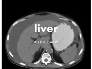
Computed tomography(1) The standard evaluation method for stable p’t Performed with Dilute water soluble oral contrast agent and intravenous contrast
Non-operative management 86% of liver injuries stopped bleeding by the time of surgical exploration 67% of operations performed are nontherapeutic Standard method of pediatric p’t and many adults
Non-operative management Criteria --hemodynamically stable --simple hepatic parenchyma laceration of inrahepatic hematoma --absence of active hemorrhage --hemoperitoneum of less than 500ml --limited need for liver related blood transfusions (12U) --absence of peritoneal sign --absence of other peritoneal injuries that would otherwise require an operation
Non-operative management Criteria --good quality CT scans --experienced radiologist --intensive care setting Currently believe that ultimate decisive factor should be the hemodynamic stability at presentation or after initial resuscitation
Non-operative management Abdominal CT --no alteration in management is indicated unless there is change in patient’s clinical course Resumption of normal activities --avoid delayed hemorrhage --avoid contact sports or heavy physical activity for 8 wks after liver injury of grades III-VI (3wks-6months)
Non-operative management Role of interventional radiology in blunt liver injury --to document active haemorrhage in subcapsular haematomas, --as a salvage alternative to surgery in the face of continuous haemorrhage in patients who remain haemodynamically stable --in the diagnosis and treatment of haemobilia --in the treatment of retained collections or perihepatic sepsis (using percutaneous techniques).
Non-operative management --Arteriography is useful in selected patients after operative perihepatic packing who have postoperative evidence of ongoing haemorrhage. --Biliary endoscopy may be helpful in the diagnosis and treatment of complications secondary to complex liver injury Complication --delayed hemorrhage, biliary fistula and liver abscess, hemobilia and bilhemia, extrahepatic bile duct stricture
Non-operative management Morbidity and death --the incidence of associated abdominal injury ranges from 13% to 35% --the incidence of truly missed injury ranges from 0.5% to 12% --the incidence of missed injury was 0.2% when strict guidelines for conservative treatment were followed and CT was used routinely. --Wrong interpretation and poor quality of the initial scan is the most common cause --A radiologist and the attending trauma surgeon read the initial CT scan, which must be of excellent quality were recommended
Non-operative management Complication --Delayed hemorrhage ‧most common, usual indication for a delayed operation ‧under strict guidelines, the incidence ranges from 0-5%, and blood transfusions were required in fewer than 20% ‧ common errors:(1)assuming that the hemorrhage is not related to the liver (2)multiple(more than four)blood transfusions in the hope that it will stop (3)misreading CT and underestimating hemoperitoneum and active bleeding
Non-operative management Complication --biliary fistula and liver abscess --Hemobilia ‧1%,iatrogenic causes most common ‧injury causes communication between the biliary tract and blood vessels ‧abdominal trauma, jaundice, RUQ colicky pain and blood in vomitus or stool point to this diagnosis ‧managed by percutaneous selective hepatic a. embolization or surgical intervention
Non-operative management Complication --bilihemia ‧rare complication of severe decelerationon injury, in which the hepatic venules and the intrahepatic bile ducts rupture ‧excessive bilirubin level ‧endoscopic sphincterotomy and biliary endostenting --Extrahepatic bile duct stricture ‧ the incidence is higher than the past ‧no uniformity of treatment criteria
Non-operative management Mortality rate --7-13% with most resulting from associated injuries --0-0.4% resulting from liver itself
Non-operative managementindications In haemodynamically stable patients with blunt hepatic injury, an expeditious abdominal CT scan haemodynamic stability rather than findings on CT determines which patients should be managed conservatively in haemodynamically stable patients less treatment is probably the best treatment most blunt hepatic injuries can be managed without operation with minimal morbidity and mortality rates
Operative management Initial control of bleeding achieved with temporary tamponade using packs, portal triad occlusion(Pringle manoeuvre), bimanual compression of the liver or even manual compression abdominal aorta above celiac trunk If hemorrhage is unaffected by portal triad occlusion(Pringle manoeuvre) by digital compression or vascular clamp, major vena cava injury or atypical vascular anatomy should be expected
Operative management Hepatotomy with direct suture ligation --using the finger fracture technique, electrocautery or an ultrasonic dissector to expose damaged vessels and hepatic duct which ligated , clipped or repaired --low incidence of rebleeding, necrosis and sepsis --effectives following blunt liver trauma requires further evaluation
Operative management Resection debridement --removal devitalized tissue --rapid compared with standard anatomical resection, which are more time consuming and remove more normal liver parenchyma --reduced risk of post-op sepsis secondary hemorrhage and bile leakage
Operative management Anatomical resection --reserved for deep laceration Perihepatic packing --Indication:coagulopathy, irreversible shock from blood loss (10u), hypothermia(32C), acidosis(PH7.2), bilobar injury,large nonexpanding hematoma, capsular avulsion, vena cava or hepatic vein injuries
Operative management Mesh rapping --new technique for grade III,IV laceration, tamponading large intrahepatic hematomas --not indicated where juxtacaval or hepatic vein injury is suspected
Operative management Omental packing Intrahepatic tamponade with penrose drains Fibrin glue Retrohepatic venous injuries --Total vascular exclusion --venovenous bypass --Atriocaval shunting --Liver transplantation
Operative management Complication --Hemorrhage,sepsis --Biliary fistula --Respiratory problems --Liver failure --Hyperpyrexia --Acalculous cholecystitis --Pancreatic, duodenal of small bowel fistula --Drainage of intra-abdominal abscess or bilioma under ultrasonography or CT guidance and embolization of AV fistula and deep bleeding vessels
Conclusion Optium results need a specialist team -experienced liver surgeon, and anaesthetist used to dealing with the coagulopathyof liver disease -interventional hepatobiliary radiologist and endoscpoist to manage post-op complication -rapid infusers, cell savers and venovenous bypass to deal with massive blood loss -Appropriate intensive care facilities -perihepatic packs to control hemorrhage -hepatotomy with direct suture ligation or resection debridement was preferred
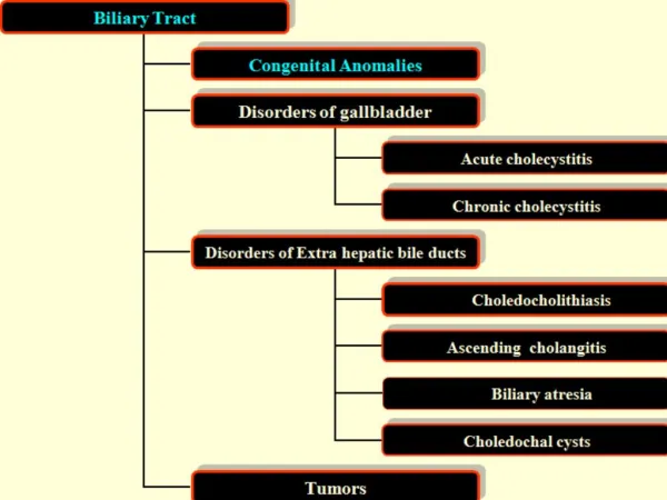
Taenia echinoccocus (cel mai frecvent) Taenia echnoccocus granularis – more severe due to exogenous vesiculation Multiple localisation: lung, brain, muscles ETHIOLOGY
Types of cysts: unic vs.multiple Structure proligerous membrane -inside the mebrane: CUTICULA is the germinative strata, producing new daughter cysts Adventiceal layer – compression and metaplazia of normal liver tiussue, forms the percyst which may impregnated with calcium salts. content–typical transparent fluid, but may become stayne dwith bile PATHOLOGY
PRETUMORAL No symptoms Alergic reactions and eosinophylia PSEUDOTUMORAL– may become evident on liver surface and produce tumoral effect on adjacent organs: - post-superior – sdr. BUDD-CHIARI - ant-inferiorduodenal compression - post-inferior–lombar expasnion may mimick renal problems, IVC compression SYMPTOMS
- centralc – may compress IVC or portal vein –portal hypertension left lobe–may compress the splenic vein: segmentary portal hypertension COMPLICATIONS SYMPTOMS
COMPLICATIONS • SISTEMIC 1. Alergic reactions – may produce even anaphylactic shock when suddenly ruptures 2. Septic complications – may become infected and behaves like liver abscess with billiary communications.
COMPLICATIONS B. LOCAL 1. Fissure: cyst may be evacuated in the billiary tree, main step to many comlpications 2. Rupture • Peritoneal cavity - usually with anaphylactic shock - may be symptom-less - during the operation Peritoneal echinoccocosis • Pleural cavity or pericardium • Bronchial tree: sudden pseudo-vomitus with salty taste, +/- alergic reactions. Risk to seed the opposite lung
TRATAMENT • No treatment- when the parasite is dead and cyst completely encapsulated • Medical treatment – antiparasitic, if the parasit may be accessed, as well as postoperative • Surgical treatment – most cases
STEPS TO FOLLOW 1.Parasite inactivation - injection in the cyst of a parasitic substance 2. Evacuation of the cyst 3. Extraction of the germinal membrane and daughter cysts 4. Manage the cavity.- care for billiary fistulas 5. LIVER RESECTION may be required
TREATMENT - alternatives • LIVER RESECTION may be required • Ideal cystectomy • Percutaneous treatment – US control • Laparoscopic treatment • Albendazole
TYPICAL EVOLUTION • Slow growth but no complications • Local complications – typical with billairy fistulas • Calcification of the membrane – associated with parasitic death