
Proteins
Proteins. General Info about Proteins. Most diverse and most important macromolecules. Our entire DNA codes for proteins only, and nothing else. Therefore they are involved in everything cells do. Protein Structure.
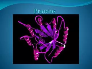

Proteins
E N D
Presentation Transcript
General Info about Proteins • Most diverse and most important macromolecules. • Our entire DNA codes for proteins only, and nothing else. Therefore they are involved in everything cells do.
Protein Structure • Proteins are polymers of amino acids folded into a specific 3D shape that belays its function. • Amino acids are made up of a central carbon atom connected to an amino group, carboxyl group, a hydrogen atom and an R group (side chain).
Amino Acids • Amino acids are considered to be amphiprotic because they possess both an acidic terminus (carboxyl group) and a basic terminus (amino group). • Humans use 20 different amino acids, so there are twenty different R-groups (shown on pages 42-43). • 8 of these amino acids are known as essential amino acids because our body cannot make them from simpler substances, so we must get these amino acids from our diet.
Amino Acids • Amino acids can be classified based on their R group in two main ways: • By polarity: polar (water-soluble) versus non-polar (lipid soluble) • By electric charge: acidic (negatively charged) versus basic (positively charged)
Linking Amino Acids to make a Protein • All proteins have a specific shape, unique to that protein. This final shape is known as the conformation of the protein and it depends entirely upon the amino acid sequence the protein contains. • In protein synthesis (the making of proteins), DNA directs the ribosomes, RNA and enzymes to start linking amino acids together in a specific sequence (Unit 3).
Linking Amino Acids to make a Protein • The joining of amino acids together is another example of a dehydration synthesis reaction. The linkage between two amino acids is known as a peptide bond. • The growing polypeptide chain will always have an amino terminus (A-terminus) and a carboxyl terminus (C-terminus). • All specific proteins have the same exact polypeptide sequence (i.e. all insulin proteins contain the exact same polypeptide sequence), which is determined by DNA.
Globular Proteins • Globular proteins are composed of one or more polypeptide chains that have a rounded, spherical conformation. • Globular proteins have four levels of structure: (p. 44 and/or handouts for diagrams)
Space filling and ribbon models of lysozyme, a protein assembled from 129 amino acids.
The primary structure of a protein is the sequence of amino acids dictated by DNA.
Sickle cell anemia is due to a single amino acid substitution in the primary structure of hemoglobin
Lysozyme Coiling and folding of the amino acid chain defines a protein’s secondary structure. This secondary structure is due to hydrogen bonds between adjacent portions of the polypeptide chain.
The tertiary structure of the protein is based upon the interactions between the R groups of amino acids. As shown in the diagram below, there are four main interactions between R groups of different amino acids.
Cytochrome c. Hydrophilic side chains are shown in green and are located mainly on the surface of the protein where they come in contact with the aqueous cytoplasm. Hydrophobic side chains are shown in red are primarily located within the interior of the protein.
The association of two or more polypeptide chains to form a functional protein constitutes the quaternary structure of a protein. Collagen Hemoglobin


