Cytoskeleton
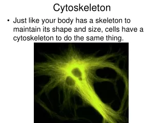
Cytoskeleton. Lecture 28 Pages 573 - 607. A cytoskeleton is needed for many many cellular functions, such as; Muscle contraction Permitting sperm to swim Immune system In fact without the functioning of the cytoskeleton life would be stagnant at the very most.

Cytoskeleton
E N D
Presentation Transcript
Cytoskeleton Lecture 28 Pages 573 - 607
A cytoskeleton is needed for many many cellular functions, such as; Muscle contraction Permitting sperm to swim Immune system In fact without the functioning of the cytoskeleton life would be stagnant at the very most. The inside of the cell is also in constant motion, and it is the cytoskeleton which provides the means to achieve this too. Mitosis & meiosis Organelle movements Cell movement The cytoskeleton is built on three types of protein filaments: Actin filamentsIntermediate filamentsMicrotubles
17_02_3 types of protein filaments Remember: AIM
Intermediate Filaments • Have the great tensile strength • Enable cells to withstand the mechanical stress associated with stretching • Called intermediate because their diameter is 10nm • Toughest and most durable of the three types • Found in the cytoplasm of most animal cells surrounding the nucleus and extending out into the cytoplasm • Found extensively at tight junctions. • Found within the nucleus too - as part of the nuclear lamina which supports and strengthens the nuclear envelope
Intermediate filament structure • Resemble rope in that many long strands are twisted together to provide tensile strength • The strands are made of elongated fibrous proteins • Each has a N-terminal globular head • Each has a C-terminal globular tail • Each has a central elongated rod domain, which is an extended alpha-helical region. These wrap around each other in a coiled-coil configuration. These are further associated non-covalently with other dimers to form tetramers. The tetramers associate together end-to-end and side-by-side also non-covalently…
The head & tail units of the fibres bind to other components. The globular domains vary greatly between different intermediate fibres
They are very evident in the axons of nerve cells which is a necessary requirement to prevent its rupture. Also present in muscle cells and epithelial cells. Shown below are the potential consequences of cells lacking these structures 17_05_strengthen_cells.jpg
The most diverse subunit family are the keratins, with different sets present in different epithelia e.g. the lining of the gut as opposed to the skin, hair, feather, claws, desmosomes 17_06_filam_categories.jpg
17_07_Plectin.jpg The intermediate filaments are stabilized by accessory proteins that transverse them and links them to other cellular structures. One important type is plectin.
Application: Nuclear enveolpe The nuclear membrane come and goes as the cell performs the cell cycle.
17_08_nuclear envelope.jpg The nuclear envelope is supported by a meshwork of intermediate filaments which are formed from lamins (not to be confused with laminin that is the extracellular matrix protein. Interestingly, the assembly and disassembly of the lamina is controlled by dephosphorylation and phosphorylation, respectively, of the lamins by protein kinases, i.e. during mitosis
Microtubles • Microtubles have a crucial role in all eucaryotic cells • Long stiff hollow tubes of protein that disassemble and reassemble to various locations in the cell. • In most cells they tend to grow from a central location within the cell called the centrosome. [it should be very clear now that the spelling of names in biology is very important - centriole, centromere, centrosome, etc.] • They extend out towards the cell periphery • They create a system of tracks across the cell • Vesicles, organelles, and other cell components travel like trains along these tracks • Keeping things in their places too, such as organelles
17_09_Microtubules.jpg The microtubles reform as the mitotic spindle during cell division along which the chromosomes move. They also form the core of the cilia and flagella that beat rhythmically.
Microtubule structure • Microtubules are built from subunits - tubulin • Tubulin is made of two subunits - alpha-tubulin and beta-tubulin bound together by ionic bonds. • These subunits stack together into long fibers microtubles • These fibers from a cylinder made of 13 parallel microtubles - each is called a protofilament • These protofilaments have a polarity • at one end there are just free alpha-tubulins - this is known as the minus end • at the other beta-tubulins - this is knowna s the plus end • this directionality is akin to defining one way streets along which material is transported…
Centrosome • Microtubules are formed by outgrowth from the specialized organizing center which controls • The number of microtubules formed • The location of these mictotubules • The orientation of these microtubules • It is known as the centrosome • It is typical present on one side of the nucleus • It is a complex structure composed of gamma-tubulin subunits • It does contain the centrioles (which are not involved in microtubule formation • Alpha/Beta subunits grow from it…
17_12_grows_shrinks.jpg The microtubules grow and contract in a dynamic fashion. This is known as dynamic instability and use a great deal of ATP
17_15_nerve_cell_axon.jpg Microtubules carry cargo along their lengths, e.g. axons of nerves It is estimated that they move material at a rate of 10 cm per day
Motor Proteins • Microtubules have a lot of associated proteins • One class are the motor proteins which bind to both actin and intermediate filaments • They use the energy of ATP hydrolysis to travel along the filaments (for both actin and intermediate) • They attach with their globular end to the filament • They attach their other end to the cargo they carry • Dozens have been identified • THEY DIFFER IN THE CARGO THEY BIND AND THE DIRECTION THEY TRAVEL • Kinesins move towards the plus end • Dyneins move towards the minus end…
Cilia and flagella contain stable microtubules moved by dynein. Cilia are small outgrowths on the cell surface that beat to move fluid across the cell - mucus in the lungs Flagella are longer structures that propel the entire cell. 17_24_cilia.jpg
The microtubules of these two structures are slightly different in that they have a ‘9 + 2’ arrangement.
Actin Filaments • Found in all eukaryotic cells • Essential for movement - cell crawling, dividing into two. • Unstable dynamic structures • Also able to bind with a whole bunch of alternative actin-binding proteins to allow them to serve a variety of functions • Act like the internal muscles of the cell - pulling the cell into various shapes • They are thin and flexible - about 7 nm in diameter • Each is a twisted chain of identical globular actin molecules - all pointing in the same direction - so they have a plus and minus side also. • Much longer than microfilaments in total length as there are many more subunits of actin in the cell than microfilament subunits (x 30 times)
17_38_myosin_I.jpg All actin-dependent motor proteins belong to the myosin family - which use ATP hydrolysis to generate movement along the filaments
17_40_Myosin_II.jpg Muscle belongs to the myosin-II subfamily of myosin - these have two ATPase heads. Clusters of these bind together to make the myosin filament