
Basics of Nervous System: Organs, Neurons, Functions
E N D
Presentation Transcript
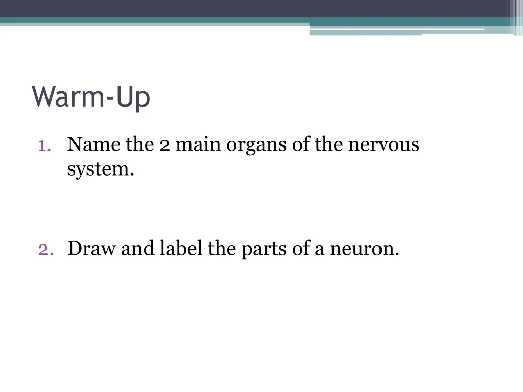
Warm-Up • Name the 2 main organs of the nervous system. • Draw and label the parts of a neuron.
Warm-Up • Label the parts of the neuron below. A D C B G F E
Warm-Up • One neuron transmits a nerve impulse at 40 m/s. Another conducts at the rate of 1 m/s. Which neuron has a myelinated axon? • List the following in order: • K+ channels open and K+ floods out of cell • Membrane is polarized (resting potential) • Neurotransmitters are released from vesicles into synaptic cleft • Na+ channels open and Na+ floods into cell • Stimulus triggers membrane depolarization • What restores the resting potential of a neuron?
Nervous System • Master controlling and communicating system
Basic Functions • Sensory input – gather information • Integration– process and interpret sensory input • Motor output – response by muscles and glands
Organization • Central Nervous System (CNS) • Brain & spinal cord • Integrative and control centers • Peripheral Nervous System (PNS) • Nerves (spinal nerves, cranial nerves) • Communication lines between CNS and rest of body • Two Divisions: • Sensory (afferent) Division: Sensory receptors CNS • Motor (efferent) Division: CNS effectors (muscles & glands)
Motor Division • Somatic nervous system (voluntary) – control skeletal muscles • Autonomic nervous system(ANS) (involuntary) – regulate smooth muscles, cardiac, glands • Subdivisions: sympathetic & parasympathetic
Nervous Tissue • Neurons(nerve cells) - transmit message Anatomy: • Cell body– contains nucleus; metabolic center • Dendrite– fiber that conveys messages toward cell body • Axon– conduct nerve impulses away from the cell body • Axon terminals– end of axon; contain neurotransmitters & release them • Synaptic cleft/synapse– gap between neurons
Nervous Tissue • Supporting cells (Neuroglia) CNS: astrocytes, microglia, ependymal cells, oligodendrocytes • barrier between capillaries and neurons • protect neurons • immune/defense • line brain and spinal cord cavities • wrap nerve fibers • produces myelin sheaths (covering) PNS: Schwann cells, satellite cells • surround large neurons • protect & cushion
Myelin: whitish, fatty material that covers nerve fibers to speed up nerve impulses • Schwann cells: surround axons and form myelin sheath • Myelin sheath: tight coil of wrapped membranes • Nodes of Ranvier: gaps between Schwann cells
Ganglia: collections of cell bodies • Bundles of nerve fibers = tracts(CNS) or nerves(PNS) • White matter: dense collections of myelinated fibers • Gray matter: unmyelinated fibers & cell bodies
Functional Classification: direction nerve impulse is traveling
Structural Classification: # processes extending from cell body
Neuron Function • Irritability: ability to respond to stimulus & convert to nerve impulse • Conductivity: transmit impulse to other neurons, muscles, or glands
Exciting a Neuron: • Cell membrane at rest = polarized • Na+ outside cell, K+ inside cell • Inside is (-) compared to outside • Stimulus excited neuron (Na+ rushes in) becomes depolarized • Depolarization activates neuron to transmit an action potential (nerve impulse) • All-or-none response • Impulse conducts down entire axon • K+ diffuses out repolarizationof membrane • Na+/K+ ion concentrations restored by sodium-potassium pump (uses ATP)
Saltatory conduction: electrical signal jumps from node to node along myelinated axon (30x faster!)
Multiple Sclerosis (MS) • Autoimmune disease • Myelin sheaths destroyed reduced to hardened lesions (scleroses) • Blindness, muscle weakness, speech disturbance, urinary incontinence • Treatment: interferons, glatiramer (hold off attacks)
Nerve Conduction • Action potential reaches axon terminal vesicles release neurotransmitters (NT) into synaptic cleft • NT diffuse across synapse bind to receptors of next neuron • Transmission of a nerve impulse = electrochemical event
Neurotransmitters • 50+ identified • Excitatory: cause depolarization • Inhibitory: reduce ability to cause action potential • Eg. acetylcholine, serotonin, endorphins
Neurotransmitters Neurotransmitter Action Affected by: Acetylcholine muscle contraction botulism, curare (paralytic), nicotine Dopamine “feeling good” cocaine, amphetamines Serotonin sleep, appetite, nausea, mood, migraines Prozac, LSD, ecstasy Endorphins inhibit pain morphine, heroin, methadone GABA main inhibitory NT alcohol, Valium, barbiturates
Reflexes • Rapid, predictable, involuntary responses to stimuli • Somatic Reflexes: stimulate skeletal muscles • Eg. jerking away hand from hot object • Autonomic Reflexes: regulate smooth muscles, heart, glands • Eg. salivation, digestion, blood pressure, sweating
Reflex Arc (neural pathway) Five elements: • Receptor – reacts to stimulus • Sensory neuron • CNS integration center • Motor neuron • Effector organ – muscle or gland
Reflex Activities Patellar (Knee-jerk) Reflex Pupillary Reflex
Patellar (Knee-jerk) Reflex Pupillary Reflex • Stretch reflex • Tapping patellar ligament causes quadriceps to contract knee extends • Help maintain muscle tone, posture, & balance • Optic nerve brain stem muscles constrict pupil • Useful for checking brain stem function and drug use
Flexor (withdrawal) reflex: painful stimulus withdrawal of threatened body part • Pin prick Plantar reflex: draw object down sole of foot curling of toes • Babinski’s sign: check to see if motor cortex or corticospinal tract is damaged
Voluntary Reactions • More neurons and synapses are involved longer response times Reflex = Involuntary Reaction Voluntary Reaction
Warm-Up • List and describe the 5 elements of a reflex arc. • List an example of a reflex. • What is the difference between a reflex and a voluntary reaction?
4 Major Regions • Cerebral Hemispheres • Diencephalon • Brain stem • Cerebellum