Comprehensive Care for Burn Patients: Guidelines and Management Strategies
This guide outlines essential information and best practices in the management of burn patients. It highlights critical burn injury statistics, the anatomy of burn wounds, assessment methods, and initial management steps. Understanding the severity of burns—including superficial, partial thickness, and full thickness—is crucial for effective treatment. The guide emphasizes airway management, fluid resuscitation, and indicators for referral to specialized burn centers. With a focus on preventing complications, this resource aids healthcare professionals in delivering optimal care to burn victims.

Comprehensive Care for Burn Patients: Guidelines and Management Strategies
E N D
Presentation Transcript
Burns Caring for the Burn Patient Victoria Siegel, RN, CNS, MSN Margarett Alexandre, RN,MS,CNA
Burn Injury Statistics • Over 2 million burn injuries and 7,000-9,000 deaths as a result of fire and burns each yr. in the U.S. • The home is frequently where burn injuries occur. • The very young and the elderly are at greatest risk for burn injuries. • Infants and toddlers are especially prone to scald injuries.http://www.ameriburn.org/
Burn Injury Statistics • School age children may incur injury as a result of playing with matches. • Teenage boys have a high incidence of electrical injuries. • Males are more common than females to be injured by burns. • 6% of burn center admissions do not survive.
Anatomy- Normal Skin Functions • Maintain fluid and electrolyte balance • Protective barrier • Regulation of temperature • Sensory functions • Immunologic functions
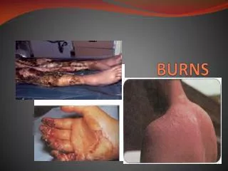
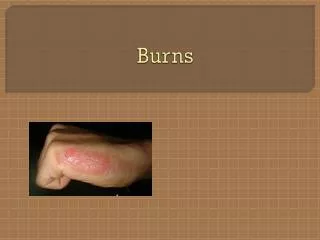
Anatomy of the burn wound • Superficial Thickness • Epidermis only portion affected • Erythema, mild edema, pain • Peeling dead skin 2-3 days after burn
Partial thickness • Partial Thickness: epidermis + partial dermis • - sparing of significant portion of hair follicles, sebaceous and sweat glands + significant portion of dermis. • Blister formation
Deep partial thickness • Second degree- Deep- destruction of large portions of hair follicles, sebaceous and substantial portion of dermis. No blisters
Full thickness burns • Full Thickness:Third degree burns- entire epidermal layers. Skin grafts required. • Escharotomies – to relieve pressure • Healing takes weeks to many months.
Deep full thickness • Fourth degree burns- underlying fascia • Damage to muscle and bones, tendons- exposed. • Sensation absent. • Wound blackened and depressed.
Severity of burn related to: • Depth • Extent • Age • Parts of body burned • Past Medical History • Concomitant injuries and illness • Presence of inhalation injury
Initial management • Goal: Limit extent of injury • Stop the burning process • Assess type of burn • Assure adequacy of ventilation and oxygenation. • Initiate restoration of hemodynamic stability. • Look for other traumatic injuries • Burn wound last priority.
Primary Survey Methodology of ABCDEF: • A- Airway/C- spine immobilization • B- Breathing • C- Circulation, cardiac status. • D- disability, neurologic deficit • E- Expose and examine • F- Fluid resuscitation
Secondary Survey • AMPLE: • A- allergies? • M- Medications/alcohol/drugs used? • P- Previous illness; PMH, last tetanus? • L- Last meal or drink • E- Events preceding injury (cause of burn?, injury occur in a closed space?, chemicals involved? Related trauma?
Respiratory tract injury • Carbonaceous sputum • Facial burns, singed nasal hairs • Agitation, tachypnea, anxiety, stupor, cyanosis, other signs hypoxemia. • Rapid resp. rate, flaring nostrils, intercostal retractions. • Hoarse voice, brassy cough, grunting or guttural respiratory sounds. • Rales, rhonchi or distant breath sounds
Airway management • Administer O2- Give 100% oxygen to all patients with burns of 20% or more TBSA. • Give 100% O2 by mask to any patient suspected of CO +/or inhalation injury. • Endotracheal Intubation- Transnasal intubation if possible, transorally if necessary. • Obtain blood gases and carboxyhemoglobin levels ASAP.
Burns • Smoke inhalation • Carbon monoxide poisoning • Assess blood gas, chest x-ray. • Listen for hoarseness and crackles • Prepare for bronchoscopy and/or possible intubation or tracheostomy for facial burns
Airway Management • Carbon Monoxide Poisoniong- 100% O2 until carboxyhemoglobin <15% • Transfer to a Burn Unit. • Inhalation injury above or below glottis- intubate immediately, suctioning, relieve dyspnea. • Circumferential burns of chest may require escharotomies.
Eschar • Necrotic tissue resulting from a burn wound. • Separates slowly from underlying viable tissue. • Good medium for microorganisms. • Failure to treat can lead to infection • Escharotomies are commonly performed.
ABA Referral Criteria Refer patients to a Burn Center: • Partial thickness burns greater than 10% total body surface area (TBSA). • Burns that involve face, hands, feet, genitalia, perineum, or major joints. • Third degree burns in any age group. • Patients with pre-existing medical disorders or trauma. • Patients requiring special social, emotional, rehab intervention
Initial treatment in ER • Establish airway • Initiate IV therapy, weigh pt. • Insert foley – hourly assessment of u/o • Insert NG tube to remove contents. • Insert CVP – hemodynamics • Baseline mental status • Initiate treatment of burn wounds • Initiate tetanus prophylaxis. • Perform a head to toe assessment
Specific Management • Flame Burns Smother the flames Remove smoldering clothing and all metal objects • Chemical Burns Brush off all chemicals present on the skin or clothing. Remove the clients clothing Ascertain the type of chemical causing the burn (acid or alkalai)
Electrical Burn At the scene, separate the client from the electrical current Smother any flames that are present Initiate cardiopulmonary resuscitation Obtain an electrocardiogram • Radiation Burns Remove the client form the radiation source If the client has been exposed to radiation from an unsealed source, remove clothing (using lead protective gloves)
Burns • Determine extent of body surface burned. • Rule of nines body divided into groups equal to about 9% of BSA. • Palm method- rough estimate adult palm is equal to 0.5% to 1% of BSA. • Lund- Browder classification- each section of body has own % according to age of pt. • Computerized mechanism in some burn units.
Fluid Resuscitation • Systemic Response: • Marked increase in peripheral vascular resistance • Reduced cardiac output- edema forms in burn injury area; blood volume decreases. • Cellular response: Full thickness burn; protein coagulation causes cell death with thrombosis of small vessels and nerve necrosis. • Goal is to maintain vital organ function and avoid complications of inadequate or excessive therapy
Parkland (Baxter) Fluid Resuscitation • Calculation of fluids for 1st 24 hrs: • Adults: Ringer’s Lactate 4ml/kg body weight x % TBSA burn. • Children: Ringer’s Lactate 4 ml/kg body weight x % burned. • Infusion rate is regulated so 50% of estimated volume is administered in the first 8 hours post burn. • Remaining 50% administered over next 16 hrs.
Fluid Resuscitation Response • Monitoring of Response- Hourly urine output. • Adults: 0.5 – 1.0 mL/kg/hr • Children: 1.0 mL/kg/hr. • Fluid and electrolytes • Weigh patient daily • Monitor vital signs, assess lung sounds.
Burns • NURSING DIAGNOSIS Impaired gas exchange decreased cardiac output Inadequate tissue perfusion Fluid volume deficit or fluid volume overload Impaired skin integrity. Risk for infection
Burns • 1. Emergent period 24-48 hours, vascular changes, shock, respiratory failure • 2. Acute phase- until all wounds heal (up to several months). Risk- infection. • 3. Rehabilitation phase- regain or compensate for loss- many years.
Initial management of burn wound • Cool the wound within 30 minutes to limit tissue damage and reduce edema but avoid excessive cooling. • Maintain blisters intact • Cover wound with clean, dry, occlusive dressing (sterile if possible). • Apply topical antimicrobial ointment if transfer to burn unit is to be delayed.
Burns • Cleanse wounds daily • Debride eschar, dress wounds, • Fine mesh gauze on granulating, healing wounds. • Promote healing to donor sites- open to air 24 hrs. post-op.
Burns • Desire normal body temp- do not expose wounds unnecessarily. • Warm ambient temp. • Warm dressing and solution to body temp. • Administer antipyretics as needed.
Burns Avoid infection Monitor for sepsis Hand hygiene Sterile dressing changes Use barrier garments Administer antibiotics
Treatment methods for burns • Method –open exposure • Burned area cleansed and exposed to air, no clothing or bedclothes over area. • Cradle over bed. • Isolation technique • Sterile linen • Room temp. 85 degrees, humidity- 40-50%
Treatment methods for burns • Method- closed • Burned area cleansed • Dressings applied and changed one to five times a day. • Standard dressing- topical antibiotics on wound, then sterile multiple gauze layers.
Treatment method for burns • Method – hydrotherapy • Place pt. in hydrotherapy tub for 20-30 min, 2X per day. • Attendants wear gowns, gloves until wounds are healed. • Tub room kept 80-90 to prevent chilling.
Wound debridement 1. Mechanical- hydrotherapy, tub, shower, forceps to remove loose, nonviable tissues 2. Enzymatic- naturally- by autolysis, spontaneous disintegration of tissues (own cellular enzymes. • Travase (sutilains)- proteolytic agent applied 3. Surgical (within first 5 days after injury) excise burn wound, then cover with skin graft or temporary covering- reduces # hydrotherapy treatments, risk- massive blood loss.
Topical medications • Silvadene-broad antimicrobial activity, no electrolyte imbalances, can cause leukopenia. • Sulfamylon- broad, used partial and full thickness, side effects- met acidosis, causes severe pain when applied. • Silver nitrate solution- broad, applied with wet, bulky dsg., restricts mobility, causes elec. imbalances, stings when applied.
Agents used in burns • Dakin’s- dress wounds that are “soupy”, aids in debridement, may inhibit clotting, causes elec. imbalances. • Betadine- may control candida, may cause elec imbalances. • Furacin- antimicrobial- effective staph aureus, may cause contact dermatitis, renal problems if burns are extensive.
Skin grafts • Biologic- viable tissue on once living tissue • To promote re-epithelialization of deep second degree burns. • To cover a wound temporarily after wound excision. • To protect granulation tissue between autografts. • Heterograft- xenograft, skin from another species (pig), • Rejection after 24-72 hours.
Skin grafts • Homograft (allograft)- • From another human (cadaver usually) • Rejection after 24 hours. • Amniotic membrane- disintegrates 48 hrs. • Artificial skin- gradually dissolves. • Autograft- first debride, then transplant • Transcyte grown in lab from foreskins.
Pressure dressings • After graft heals • Prevents formation of contractures and tight hypertrophic scars • Uniform pressure over burned surfaces. • Worn 23 hrs. a day.
Burns- body positions • Encourage prone and supine positions for a definite interval each day. • Frequent position changes • Burns on neck and chin- encourage position of neck hyperextension for part of the day. • Burns on hand- consult M.D. for specifics.
Burns- preventing mobility limitations • Contractions – serious complication. • Help to maintain range of joint motion • Exercises to prevent and correct contractures are begun ASAP- stable • PT/OT, Hubbard tank • Chewing gum and blowing up balloon – prevent facial contractures.
Burns- Pain Management • Provide analgesic medication 30 minutes prior to painful treatments. • Provide clear explanations to gain patient’s cooperation. • Handle burned parts gently. • Use careful sterile technique (infection causes more pain).
Burns: Pain Management • PCA, imagery , breathing techniques, enhance coping strategies. • Pt. and family education and support • Patient may need years of PT and OT. • Psych support for trauma suffered and body image changes endured.
Burns – emotional responses • Patient response- aggression • Nursing approach: • Acknowledge ability to cope. • Provide structure; allow pt. choices when possible. Pt. needs some control. • Burn team must be sensitive to emotional and psychological needs of patient and family.
Emotional responses • Depression- • Nursing approach- support patient, listen. • Encourage verbalization of frustrations. • Paranoia- • Nursing approach- acknowledge c/o fear. • Investigate all complaints. • Support pt. • Provide reality orientation.
Teaching and Discharge Instructions • Care of the healed burn wound • Nutritional needs • Prevention of injury • Recognition of S&S of complications. • Methods of re-socialization. • Evaluation- Any signs of infection?, Diet being followed?, Pt. involved? Pt. understand D/C instructions?
NCLEX TIMEBlisters are a classic sign of which classification of burn? • 1. Superficial • 2. Superficial partial thickness • 3. Deep partial thickness • 4. Full thickness