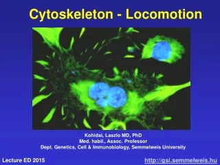
Cytoskeleton - Locomotion
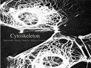
Cytoskeleton - Locomotion. Kohidai, Laszlo MD, PhD Med. habil., Assoc. Professor Dept. Genetics, Cell & Immunobiology, Semmelweis University. 2008. Main functions of cytoskeleton. Determines the shape of the cell Anchores organelles Movement of organelles Tensile strength

Cytoskeleton - Locomotion
E N D
Presentation Transcript
Cytoskeleton - Locomotion Kohidai, Laszlo MD, PhD Med. habil., Assoc. Professor Dept. Genetics, Cell & Immunobiology, Semmelweis University 2008
Main functions of cytoskeleton • Determines the shape of the cell • Anchores organelles • Movement of organelles • Tensile strength • Movement of chromosomes • Polarity • Motility
Cytoskeleton • Microfilaments (actin) • Microtubuli (tubulin) • Intermedier filaments • Microtubule associated proteins (MAP-s) • Motor proteins
Microfilaments Microtubuli Intermedier filaments
SLIDING Globular proteins Ca2+ ATP Motor proteins Fibrillar proteins
Polymerization of actin + ATP ADP Depolymerization - cytochalasin – inh. phalloidin - stabilizer ATP ADP Pi Polymerization - slow
Actin - still in Prokaryots ! ((Ent et al. Nature 2001,413, 39)
Moving cytoplasm Stationary (cortical) cytoplasm Plasma membrane Actin filaments Cell-wall Chloroplasts Cyclosis • Transitional connections between actin and myosin • Ca2+, temperature- and pH-dependent (Lodish, H. et al. Mol. Cell Biol. 2000, 767)
„Fountain” mechanism Ca2+-dep. requires ATP Mono- Poly- Lobo- podial Filo- Reticulo- Formation of pseudopodium stress-fibrillums integrins
Cross-linking proteins of actin contractile bundle a actinin – in stress fibr. gel-like network filamin - cortex „tight” parallel bundle fimbrin – in filopodium
Migrating keratinocyte 15 mm/sec Formation of lobopodium microtubuli actin-network
- + Regulator proteins of actin polymerisation gCAP39 Severin Gelsolin Villin CapZ Tropomodulin Cofilin Severin Gelsolin
Actin polymerisation – acrosomal-reaction (Lodish, H. et al. Mol. Cell Biol. 2000, 767)
local actin polymerization • speed: 10 mm/min • high ability to transmit • in tissues Listeria monocytogenes actin (Fred Soo & Julie TheriotLaboratory)
Model of actin nucleation WASP = Wiscott-Aldrich syndr. prot.
Structure of cortical region (Svitkina, TM, Borisy GG J. Cell Biol. 1999, 145, 1009)
Myozin I. Arp2/3 Profilin - G-aktin Filamin Integrin Actin – membrane links membrane F-Actin
Profilin-mechanism Tb4 =timozin b4 Proline-rich protein (Lodish, H. et al. Mol. Cell Biol. 2000, 767)
Filamin – Membrane link filamin actin
Structure of focal contact actin filament a actinin vinculin + paxillin talin integrin fibronectin
Thrombocyte Glycophorin Ankyrin Spectrin tetramer Muscle Epithel A plasma membrane – cortex links ((Lux SE, 1979 Nature 281:426)
E Electromagnetic field induces the transformation of cytoskeleton and formation of pseudopodia Adhesion plaque + + + - -
ATP - ADP Pi Myosin head Ca2+-dependent phosphorylation and its effect on the 3D strcture light chain heavy chain a helix myosin I. 150 kD monomer myosin I I. 260 kD Head: - ATP-ase - motor dimer
Distribution of myosines in the migrating Dyctiosteliumand in dividing cell myozin I. (green) myozin II. (red) (Fukui, Y. Mol. Cell Biol 2000, 785))
+ - Main types of interactions between the globular and fibrillar components of cytoskeleton membrane
MT-blocked F-actin blocked Non-treated
Tubulin – still in Prokaryotes ! FtsZ Tubulin (Margolin Laboratory, University of Texas)
Polymerization of tubulin GTP Polymerization - fast GTP GTP GTP Protofilamentum (strait) GDP GDP GDP GDP Protofilamentum (curved) Depolymerization
Nucleation Elongation Dynamics of microtubule-assembly - + incorporation balanced release
Role of g-tubulin in nucleation (Wiease et al. Curr.Opin.Struct.Biol. 1999, 9, 250)
Interphase cell centrosome Cilla Basal body Dividing cell spindle Neuron centrosome axon Microtubular systems in the cells -Centrosome - Cilia / flagellum - Mitotic system - Vesicular transport
specialized region of the cortex MTOC = Mikrotubul organizing center g-tubulin ((Brinkley, B.R. Encyclop. Neurosci. 1987, 665)
24 nm abdimer Protofilaments atubulin btubulin Network of microtubuli Fibroblast
Cilia cilia flagellum Paramecium
tubulin (13 ill. 11 protofilaments) A B dynein-arms nexin
The arm moves toward the - pole Composition of dynein-arms ATP-independent binding ATP-dependent hydrolisis
The role of dynein arms in beating of cilia Bending „Telescoping” Proteolysis
Molecules composing the cilia more than 250 types of molecules • 70% a and b tubulin • dynein arms • outer - 9 polypeptides - ATP-ase • inner – composition varies • radial spokes - 17 polypeptides
intermedier filament i.e. vimentin microtubule = rupture actin filament Mechanical characterization of cytoskeleton components deformation force
Role of intermedier filaments Buffer against external mechanical stress Tissue specificity Nucleus – lamines (lamina fibrosa) Epithel – keratin Connective tissue Muscles Neuroglia Neurones - neurofilaments }vimentin
Structure of intermedier filamentums (Lodish, H. et al. Mol. Cell Biol. 2000, 767)
Domain structures of intermedier filamentums H2N- a helical domain -COOH keratins vimentin neurofilam. prot. nuclear prot
Intermedier filaments Keratin filaments Vimentin-like filaments ! They DO NOT co-polymerise !
Microvilli myosin I. actin villin „terminal web” • a rigidbundle composed by20-30 actin mol.s • actin + on the apical part • villin is the linker molecule of actins • „terminal web” = intermed.fil. + spectrin • myosin I. and calmodulin anchore to the surface membrane
SEM structure of microvilli actin bundle linker molecules „terminal web”