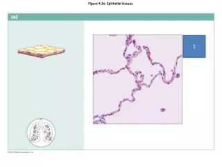
Epithelial Tissues
Epithelial Tissues. 4 Types of Tissue. Epithelial Connective Muscle Neural Can you come up with 2-3 basic functions for each of these?. Characteristics of Epithelia. Predominantly made of cells all cells tightly bound by cell junctions Polarity ( apical and basal surfaces)

Epithelial Tissues
E N D
Presentation Transcript
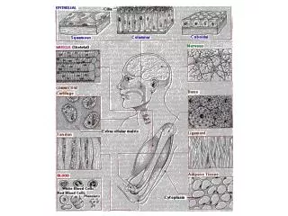
4 Types of Tissue • Epithelial • Connective • Muscle • Neural • Can you come up with 2-3 basic functions for each of these?
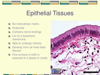
Characteristics of Epithelia • Predominantly made of cells • all cells tightly bound by cell junctions • Polarity (apical and basal surfaces) • Attached to basallamina • Avascular (lack blood vessels) • Regenerate via stem cells
Where is Epithelia found? • Covers exposed surfaces • Skin, digestive tube, reproductive tract, ureter, bladder, trachea, lungs • Lines internal passageways • Blood vessels, lymph system • Forms glands • Sebaceous (mammary), salivary, gastric, intestinal, mucous
Functions of Epithelia • Provide Physical Protection • Control cell permeability • Provide sensation - neuroepithelia • Produce secretions via glands
Functions of Epithelia • Physical Protection • Protect all surfaces exposed to the environment (external & internal) • Mechanical (abrasion) • Biological & chemical (bacteria, viruses & their byproducts) • Dehydration
Functions of Epithelia • Control cell permeability – absorption • Anything entering or leaving body crosses epithelia • Sensitive to various stimuli • PTH - increase Ca2+ absorption across epithelial cells of small intestine • Aldosterone - increases Na+ absorption across epithelia of LOH • Vitamin C – promotes iron absorption in small intestine
Functions of Epithelia • Provide sensation - neuroepithelia • Large sensory nerve supply • mechano, electro, chemoreceptors • All special senses provided by specialized epithelia
Functions of Epithelia • Form glands (glandular epithelia) that produce secretions • Exocrine - released onto epithelial surface via ducts • sweat, tears, milk • Endocrine - released into blood or lymph for action elsewhere • Hormones • ductless • What is the difference between exocrine and endocrine glands & secretions? Where and how they DELIVER those secretions
Specialized for different functions • Move fluids over the epithelium • Protection & lubrication (mucus) • Move fluids through the epithelium • permeability • Produce secretions • Protection, messengers, waste removal, nutrient supply
Mechanical & Chemical Barriers • Physical integrity is maintained by: • intercellular connections • attachment to basal lamina • maintenance and repair
Intercellular Connections • Support • Communication
Large Connections • CAMs (cell adhesion molecules): • Transmembrane proteins: cell membrane-cell membrane connections • Intercellular cement: • Proteoglycans (extracellular polysaccharides linked by polypeptide chains) • Glycosaminoglycans • Hyaluronan
3 Cell Junctions • Form bonds with other cells or extracellular material: • tight junctions • gap junctions • desmosomes
Tight Junctions • Between 2 cell membranes • Adhesion belt attaches to terminal web • Prevents passage of water & its solutes • Acids • ingested Bacteria • EX: Isolates waste & metabolic byproduct throughout lumen
Desmosomes • CAMs, dense areas, and intercellular cement • Button desmosomes • Ties cells together • Supported internally by intermediate filaments; Allow bending and twisting • Hemidesmosomes • Ties cell to basal lamina
Lamina lucida(closer to ET): thin layer secreted by epithelia barrier to proteins Lamina densa (closer to CT): thick fibers Secreted by connective tissue strength and filtration Attachment to Basal Lamina
Repairing & Replacing Epithelia • Epithelial cells are replaced by division of germ cells (stem cells) • Lie just superior to basal lamina
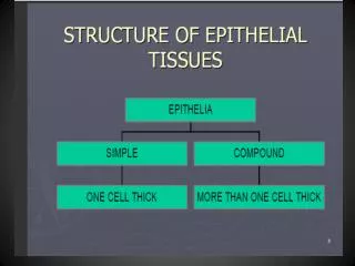
Cell Arrangements • Simple: http://a-s.clayton.edu/biology/biol1151L/lab03/lab_3.htm • Single layer of cells = fragile • Often used for filtration = secretions & absorption • Stratified • 2 or more layers of cells • Use top layer to identify (as squamous, cuboidal etc.) • Often seen in ‘wear and tear’ areas (high mechanical or chemical stress) • Pseudostratified • One layer that looks like many layers • Seen in areas needing to stretch (bladder walls)
Classes of Epithelia • Based on shape and # of layers Table 4–1
Surfaces • Apical • Basal • Basement Membrane • basal lamina: collagen, laminin, proteoglycans • Epithelial secretions • reticular lamina: reticular fibers, fibronectin, glycoproteins • connective tissue secretions
Surface Variations • Plain • Cilia • Propel substances over surfaces • Microvilli • Increase exposed surface area • Often in columnar epithelia
Modes of Glandular Secretion • What is lost during secretion? • Apocrine • Merocrine • Holocrine
Merocrine Secretion • Secretory products secreted via vesicular exocytosis • Most common form of secretion – often continuous • Ex: Pancreatic cells, sweat, saliva Figure 4–6a
Apocrine Secretion • Cytoplasm & secretion are excreted • milk & underarm persperation Figure 4–6b
Holocrine Secretion • Entire cell lost with secretion. • Stem cells divide to replace lost cells • Ex: Sebaceous glands Figure 4–6c
Glands: Arrangement of cells • Unicellular: • Goblet cells • Multicellular: • Tubular – Brunner’s gland in duodenum • Alveolar – Sebaceous/Oil glands • Combination – Salivary glands • Mucous = mucin: • A glycoprotein that combines with water
Secretory Glands: General Types • Serous • Secrete a watery substance containing enzymes • Saliva with alpha-amylase • Mucous • Secrete mucin • hydrates to form mucus • Mixed Exocrine