Download
1 / 40
410 likes | 494 Vues
Learn about the tools and techniques used in isolating and purifying DNA from various sources, such as blood, cells, bacteria, and forensic samples. Understand the steps involved, basic rules, and PCR applications.

E N D
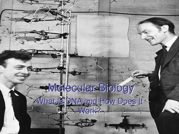
Tools used in Molecular Biology
Fig A :Basic cell types Fig B :Basic molecular process in Cell
PRINCIPLES OF DNA ISOLATION & PURIFICATION DNA can be isolated from any nucleated cell. DNA is a giant anion in solution.
Sources of DNA include Blood Buccal cells Cultured cells (plant and animal) Bacteria Biopsies Forensic samples i.e. body fluids, hair follicles, bone & teeth roots.
DNA isolation is a routine procedure to collect DNA for subsequent molecular analysis. There are three basic steps in a DNA extraction: Cell disruption:- This is commonly achieved by grinding or sonicating the sample. Removing membrane lipids by adding a detergent. Isolation of DNA:- Removing proteins by adding a protease (optional but almost always done). Precipitating the DNA :-usually ice-cold ethanol or isopropanol is used. Since DNA is insoluble in these alcohols, it will aggregate together, giving a pellet upon centrifugation. This step also removes alcohol soluble salt.
Basic rules • Blood – first lyse (explode) the red blood cells with a gentle detergent such as Triton-X-100. • Wash cells – haemoglobin (and other pigments) inhibits restriction enzymes and TAQ polymerase. • Work on ice to slow down enzymatic processes. • Wear gloves to protect your samples from you!! • Autoclave all solutions and store in fridge (except SDS and organic solvents!) • Keep all pellets & supernatants until you have the DNA you want.
Getting to the DNA Cells – lyse all cells in presence of : NaClso that DNA is stabilised and remains as a double helix, EDTA which chelates Mg++ and is a co-factor of DNAse which chews up DNA rapidly. anionic detergent SDS which disrupts the lipid layers, helps to dissolve membranes & binds positive charges of chromosomal proteins (histones) to release the DNA into the solution. Include a protease(proteinase K) to digest the proteins incubate the solution at an elevated temperature (56oC to inhibit degradation by DNAses) for 4-24 hrs.
Getting rid of the protein • Organic solvent extraction using equal volume phenol:chloroform (24:1) • Protein at the interface after centrifugation (10000 rpm at 10o c for 10 min.)
Precipitating the DNA add 2.5 - 3 volumes ice-cold 95% ethanol to the DNA & leave at -20oC overnight. Centrifuge sample at 10000 rpm ,10 min., 40C. Wash DNA pellet to remove excess salt in 70% EtOH and air-dry. Resuspend in sterile distilled water(pH7.4) Store at 4oC or frozen at -20oC long term.
Quantifying the DNA • The amount of DNA can be quantified using the formula: DNA concentration (g/ml) = OD260 x 100 (dilution factor) x 50 g/ml 1000 • Nucleic acids have a peak absorbance in the ultraviolet range at about 260 nm • 1 A260 O.D. unit for dsDNA = 50 µg/ml • 1 A260 O.D. unit for ssDNA = 33 µg/ml • 1 A260 O.D. unit for RNA = 40 µg/ml
DNA purity • The purity of the DNA is reflected in the OD260:OD 280 ratio and must be between 1.6 and 2.00. < 1.6 – protein contaminated > 2.0 – chloroform / phenol contaminated • Repurify sample.
Summary Sample for DNA extraction Lysis of cells at elevated temperature + detergent + enzyme in salt buffer Removal of cellular proteins Precipitation of nucleic acids with ethanol Quantitation and purity measurement of DNA
Gel electrophoresis The basic principle is that DNA, RNA, and proteins can all be separated by means of an electric field. In agarose gel electrophoresis, DNA and RNA can be separated on the basis of charge, size by running the DNA through an agarose gel. Proteins can be separated on the basis of size by using an SDS-PAGE gel, or on the basis of size and their electric charge by using what is known as a 2D gel electrophoresis.
Step 1 Step 2 Step 3 Step 4
Polymerase chain reaction (PCR) The polymerase chain reaction is an extremely versatile technique for copying DNA. PCR allows a single DNA sequence to be copied (millions of times), or altered in predetermined ways, means selectively amplifying a particular segment of DNA PCR has many variations, like reverse transcription PCR (RT-PCR) for amplification of RNA, and real-time PCR (QPCR) which allow for quantitative measurement of DNA or RNA molecules.
Materials need for PCR • Template DNA • Reaction buffer (Tris, ammonium ions magnesium ions) • Nucleotides (dNTPs : dATP, dGTP, dCTP, dTTP) • Primers • DNA polymerase (usually Taq)
PRIMER A primer is a strand of nucleic acid that serves as a starting point for DNA synthesis. These primers are usually short, chemically synthesized oligonucleotides, with a length of about twenty bases. They are hybredized to a target DNA, which is then copied by the polymerase. minimum primer length used in most applications is 18 nucleotides. Replication starts at the 3'-end of the primer, and copies the opposite strand. In most cases of natural DNA replication, the primer for DNA synthesis and replication is a short strand of RNA .
Steps in PCR • Denaturation (95°C), 30 sec. • annealing (55–60°C), 30 sec. • extension (72°C),time depends on product size.
PCR The process follows the principle of DNA replication
REACTION MIX REACTION ANALYSIS
Applications of PCR • A common application of PCR is the study of patterns of gene expression. • The task of DNA sequencing can also be assisted by PCR. • PCR has numerous applications to the more traditional process of DNA cloning. • An exciting application of PCR is the phylogenic analysis of DNA from ancient sources • A common application of PCR is the study of patterns of genetic mapping • PCR can also used in Parental testing, where an individual is matched with their close relatives.
Limitations • Need for target sequence information • Primer design for unexplored ones • Boundary region of DNA to be amplified must be known • Short size and limiting amounts of PCR products UPTO 5KB can be easily amplified Up to 40kb can be amplified with some modifications Can not amplify >100kb Can not be used in genome sequencing projects.
Southern blotting Southern blot is a method for probing for the presence of a specific DNA sequence within a DNA sample. DNA samples are separated by gel electrophoresis and then transferred to a membrane by blotting via capillary action. The membrane is then exposed to a labeled DNA probe that has a complement base sequence to the sequence on the DNA of interest. less commonly used due to the capacity of other techniques, such as PCR. Southern blotting are still used for some applications such as measuring transgene copy number in transgenic mice, or in the engineering of gene knockoutembryonic stem cell lines.
Northern blotting The northern blot is used to study the expression patterns of a specific type of RNA molecule as relative comparison among a set of different samples of RNA. RNA is separated based on size and is then transferred to a membrane then probed with a labeled complement of a sequence of interest. The results may be visualized through a variety of ways depending on the label used. Most result in the revelation of bands representing the sizes of the RNA detected in sample. The intensity of these bands is related to the amount of the target RNA in the samples analyzed. It is used to study when and how much gene expression is occurring by measuring how much of that RNA is present in different samples. one of the most basic tools for determining at what time, and under what conditions, certain genes are expressed in living tissues.
Western blotting • In western blotting, proteins are first separated by size, in a thin gel sandwiched between two glass plates in a technique known as SDS-PAGEsodium dodecyl sulphate polyacrylamide gel electrophoresis. • The proteins in the gel are then transferred to a nitrocellulose, nylon or other support membrane. • This membrane probed with solutions of antibodies. Antibodies specifically bind to the protein of interest & visualized by a variety of techniques, including colored products, chemiluminescence, or autoradiography. • Antibodies are labeled with enzymes. When a chemiluminescentsubstrate is exposed to the enzyme it allows detection. • Using western blotting techniques allows not only detection but also quantitative analysis.
Molecular markers Molecular marker are based on naturally occurring polymorphism in DNA sequence(i.e. base pair deletion, substitution ,addition or patterns). Genetic markers are sequences of DNA which have been traced to specific locations on the chromosomes and associated with particular traits. It can be described as a variation that can be observed. A genetic marker may be a short DNA sequence, such as a sequence surrounding a single base-pair change (single nucleotide polymorphism, SNP), or a long one, like mini satellites.
Some commonly used types of genetic markers are RFLP (or Restriction fragment length polymorphism) AFLP (or Amplified fragment length polymorphism) RAPD (or Random amplification of polymorphic DNA) VNTR (or Variable number tandem repeat) Micro satellite polymorphism, SSR (or Simple sequence repeat) SNP (or Single nucleotide polymorphism) STR (or Short tandem repeat) SFP (or Single feature polymorphism) DArT (or Diversity Arrays Technology) RAD markers (or Restriction site associated DNA markers)
There are 5 conditions that characterize a suitable molecular marker Must be polymorphic Co-dominant inheritance Randomly and frequently distributed throughout the genome Easy and cheap to detect Reproducible
Molecular markers can be used for several different applications including Germplasm characterization, Genetic diagnostics, Characterization of transformants, Study of genome Organization and phylogenic analysis. Paternity testing and the investigation of crimes. Measure the genomic response to selection in livestock
RFLP (Restriction fragment length polymorphism) RFLPs involves fragmenting a sample of DNA by a restriction enzyme, which can recognize and cut DNA wherever a specific short sequence occurs. A RFLP occurs when the length of a detected fragment varies between individuals and can be used in genetic analysis. Advantages: • Variant are co dominant • Measure variation at the level of DNA sequence, not protein sequence. Disadvantage: • Requires relatively large amount of DNA
AFLP ( Amplified fragment length polymorphism) In this analysis we can amplify restricted fragments and reduces the complexity of material to be analyzed (approx 1000 folds).it can be used for comparison b/wclosely related species only. Advantages: • Fast • Relatively inexpensive • Highly variable Disadvantage: • Markers are dominant • Presence of a band could mean the individual is either homozygous or heterozygous for the Sequence - can’t tell which?
RAPD ( Random amplification of polymorphic DNA) Random Amplification of Polymorphic DNA. It is a type of PCR reaction, but the segments of DNA that are amplified are random. Advantages: • Fast • Relatively inexpensive • Highly variable Disadvantage: • Markers are dominant • Presence of a band could mean the individual is either homozygous or heterozygous for the Sequence - can’t tell which? • Data analysis more complicated
Micro satellite polymorphism, SSR or Simple sequence repeat Microsatellites, Simple Sequence Repeats (SSRs), or Short Tandem Repeats (STRs), are repeating sequences of 1-6 base pairs of DNA. Advantages: • Highly variable • Fast evolving • Co dominant Disadvantage: • Relatively expensive and time consuming to develop
SNP A single-nucleotide polymorphism (SNP, pronounced snip) is a DNA sequence variation occurring when a single nucleotide — A, T,C, or G — in the genome (or other shared sequence) differs between members of a species or paired chromosomes in an individual. Used in biomedical research ,crop and livestock breeding programs.
STR A short tandem repeat (STR) in DNA occurs when a pattern of two or more nucleotides are repeated and the repeated sequences are directly adjacent to each other. The pattern can range in length from 2 to 16 base pairs (bp) (for example (CATG)n in a genomic region) and is typically in the non-coding intron region Used in forensic cases. used for the genetic fingerprinting of individuals
Future aspects • For agricultural development and environment protection. • To ensure food security for ever growing human population.