Download
1 / 28
290 likes | 507 Vues
Neuroimaging with MRI. Dr Mohamed El Safwany , MD. Intended learning outcome. The student should learn at the end of this lecture principles neuroimaging with MRI. Topics. Quick overview of MRI physics (all on one slide!) Some images and their applications

E N D
Neuroimaging with MRI Dr Mohamed El Safwany, MD.
Intended learning outcome The student should learn at the end of this lecture principles neuroimaging with MRI.
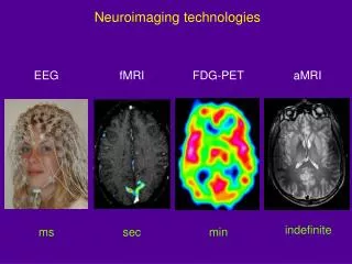
Topics • Quick overview of MRI physics (all on one slide!) • Some images and their applications • T1-weighted = gray/white/CSF delineation • T2-weighted = detection of tissue abnormalities • T2*-weighted = venography • Contrast agents • Enhancement of signals from various tissue types/conditions • Diffusion weighted imaging • Imaging brain function with MRI
MRI 1) Put subject in big magnetic field [and leave him there] Magnetizes the H nuclei in water (H2O) 2) Transmit radio waves into subject [about 3 ms] Perturbs the magnetization of the water 3) Turn off radio wave transmitter 4) Receive radio waves re-transmitted by subject’s H nuclei Manipulate re-transmission by playing with H magnetization with extra time-varying magnetic fields during this readout interval [10-100 ms] Radio waves transmitted by H nuclei are sensitive to magnetic fields — those imposed from outside and those generated inside the body: Magnetic fields generated by tissue components change the data and so will change the computed image 5) Store measured radio wave data vs time Now go back to 2) to get some more data [many times] 6) Process raw (“k-space”) radio wave data to reconstruct images
Coronal T1-weighted Image with Gadolinium Contrast Note enhancement of arteries, venous sinuses, choroid plexus, and dura mater
T1-Weighted Images • Images whose design (timing of radio pulses and data readout) is to produce contrastbetween gray matter, white matter, and CSF
Three Slices from a Volume • A single acquisition is somewhat noisy • Previous T1-weighted image was actually average of 4 separate acquisitions (to average out noise) • MRI can be a 2D or a 3D acquisition technique
Some Bad MR Images • Subject moved head during acquisition • Ghosting and ringing artifacts • Might be OK for some clinical purposes, but not much use for most quantitative brain research
T2-Weighted Images • Often better than T1-weighting in detecting tumors and infarcts (usually radiologists look at both types of scans) Same subject
T2*-Weighted Images • Designed to make venous blood (with lots of deoxy-hemoglobin) darker than normal tissue = venography Output image
MRI Contrast Agents • Chemicals injected into blood, designed to alter MRI signal by affecting magnetic environment of H nuclei • Purpose is to increase contrastof some tissue type • Most commonly used is Gd-DTPA (Magnevist) • Gadolinium ion (highly magnetizable) chelated to a molecule that won’t pass an intact blood-brain barrier • Makes T1-weighted images brighter where it accumulates and makes T2- and T2*-weighted images darker • Deoxy-hemoglobin is an endogenousT2* agent
Tumor: T2 and T1+contrast T2-weighted T1-weighted post-contrast
T2* MRV on a Seizure Patient Bad Gd-enhanced T1-weighted Gd-enhanced T2*-weighted
Diffusion Weighted Imaging • Water molecules diffuse around during the imaging readout window of 10-100 ms • Scale of motion is 1-10 microns size of cells • Imaging can be made sensitive to this random diffusive motion (images are darkened where motion is larger) • Can quantify diffusivity by taking an image without diffusion weighting and taking a separate image with diffusion weighting, then dividing the two: • Can thus compute images of ADC from multiple (2+) scans
DWI in Stroke • ADC decreases in infarcted brain tissue within minutes of the vessel blockage • Stroke damage doesn’t show up on T1- or T2-weighted images for 2-3 days post-blockage • DWI is now commonly used to assess region of damage in stroke emergencies
Diffusion Tensor Imaging • Diffusive movement of water in brain is not necessarily the same in all directions — not isotropic • Diffusion weighted MR images can be designed to give more weight to diffusion in some directions than in others • By acquiring a collection (7+) of images with different directional encodings, can compute the diffusion tensor in each voxel .
DTI Results Unweighted (baseline b=0) image Fractional Anisotropy (FA): Measures how much ADC depends on direction FA Color-coded for fiber directionality: x=Red y=Green z=Blue
Brain Activation Map Time series analysis results overlaid on T1-weighted volume
Applications of FMRI • Clinical (in individuals): • Pre-surgical mapping of eloquent cortex to help the surgeon avoid resecting viable tissue • Can combine with DTI to help surgeon avoid important white matter bundles (e.g., cortico-spinal tract) • Measure hemispheric lateralization of language prior to temporal lobe surgery for drug-resistant epilepsy
MRI is: • Widely available • Harmless to subject ifproper safety precautions are used • Very flexible: can make image intensity (contrast) sensitive to various sub-voxel structures • Still advancing in technology and applications • Still in a growth phase for brain research
Text Book David Sutton’s Radiology Clark’s Radiographic positioning and techniques
Assignment Two students will be selected for assignment.
Question Define role of diffusion imaging in acute cerebral infarction?