Download
1 / 22
250 likes | 729 Vues
Pathomechanics of hip Joint (part 1) practical section. Lecturer: Dr. Manal Radwan Salim Demonstrators: Dr.Mohammed Arafaat Dr. Haytham Essawy Dr. Atef Mohammed Dr. Mai Tolba 5 th practical section Fall 2013-2014 3-11-2013. hip/ Thigh Osteokinematics:.

E N D
Pathomechanics of hip Joint (part 1) practical section Lecturer: Dr. ManalRadwanSalim Demonstrators: Dr.MohammedArafaat Dr. HaythamEssawy Dr. Atef Mohammed Dr. Mai Tolba 5th practical section Fall 2013-2014 3-11-2013
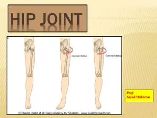
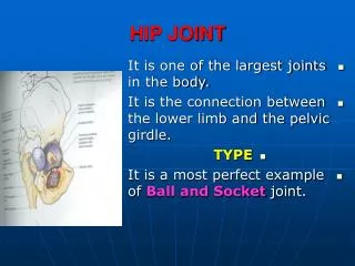
hip/ Thigh Osteokinematics: • The hip is the articulation between the large spherical head of the femur and the deep socket provided by the acetalum of pelvis, the femoral head is located jut inferior to the middle third of the inguinal ligament
1- The femur: is the longest bone of the human body. The femoral head projects medially for an articulation with the acetabulum. The femoral shaft courses slightly medial (convex medially i.e. causes the normal valgus knee in the frontal plane), thereby placing the knees and feet closer to the midline of the body.
In the sagital plane: The femoral shaft convex anteriorly (bows anteriorly) with weight bearing. In the transverse plane: The upper part of femoral shaft is externally twisted (rotated) over its distal parts (femoral condyles). so that the neck of femur is moved anteriorly with an angle of around 15 degrees in relation to femoral condyles (normal antevresion angle).
b) Angle of torsion: Normal anteversion It describes the relative rotation(twist) that exists between the shaft and neck of the femur.
Normally as viewed from above, the femoral neck longitudinal axis projects anterior to a medio-lateral(transverse) axis of the femoral condyles (10-15 degrees). Since the hip joint can only tolerate a limited amount of torsion of the head without threatening congruence.
Pathologically: • Any abnormality in the angle can change the hip joint stability via changing the location of the femoral head in the acetabulum. • n.b. greater degree of femoral antversion or retroversion may be seen distally at the femoral condyles.
Excessive Anteversion: • Any increase in the anterior angulation (excessive anteversion) > 15 degree results in a greater external rotation of femur, thus femur is less congruent in the anatomical standing position, foot neutral on ground, this causes the pelvis to be moved anteriorly in the transverse plane, or there is anterior instability of hip with possibility of anterior dislocation which actually not common to occur.
Normally this external rotation which is compensated by internal rotation of tibia, intoed position. In intoed position, the excessively antiverted head is more positioned in the acetabulum, more congruent position
Femoral retroversion: • Conversely a greater degree of external rotation. Patients who toe out may have an excessive retroversion (i.e. internal rotation of femur).
Angles of the femur: a) Neck shaft angle: (angle of inclination) • It is an angle which presents in the frontal plane between the longitudinal axis of the femoral neck and the longitudinal axis of the femoral shaft. • - The longitudinal axis of the neck : is the line which runs from the center of the head in midline of the neck to the implantation of the neck between the trochanters. • The longitudinal axis of the shaft :is the line drawn from midway of the trochanteric region to the middle of the knee joint (anatomical axis).
Its magnitude: In normal adult person: it is about 125 degrees In children, it is about 150 degrees, but by the process of weight bearing, compression of the head and neck of the femur occurs and then the neck shaft angle decreases.
Its functions: It displace the proximal shaft of the femur laterally away from the joint. Thereby reducing the likelihood of bony impingement against the pelvis. It allows more degrees of freedom of the hip by moving the longitudinal axis of the femur away from the hip bone thus allow more degree of freedom to the hip joint.
Pathologically: • If the medial angulation between the neck and the shaft increases more than 125 degrees. It is called coxavalga(bend aoutward). In this case the joint has limited ROM as the greater trochanter impinges with pelvic bones
Pathologically: • If the medial angulation between the neck and the shaft decreases less than 125 degree, it is called coxavara (bend inward). • In this case the joint has better ROM capbilities, but more load on the glutesumedius due to decreasing its moment arm of action.
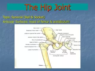
2- Acetabulum: the acetabulum projects laterally from the pelvis with a varying amount of inferior and anterior tilt. A misaligned acetabulum doe not adequately cover the femoral head, often causing chronic dislocation and osteoarthritis. Two angles describe the extent to which the shape of the acetabulum naturally covers the femoral head:
Angles of acetabulum: A-Center Edge angle" CEA“: is an angle between two lines: First line connects the lateral rim of the actabulum and the center of the femoral head. Second line is a vertical line. Its functions: *It determines the amount of inferior tilting of the acetabulum. *It describes the extent to which the acetabulum covers the femoral head within the frontal plane i.e. the normal center edge angle provides a protective shelf over the femoral head.
Magnitude: It is highly variable on average measures about 22 to 42 degrees in the x-ray of adults. A smaller CEA of the acetabulum may result in diminished coverage of the head of the femur and an increased risk of superior dislocation.
Pathologically: *a more vertical alignment (i.e., a smaller angle) offers less containment of the femoral head and is associated with an increased risk of superior dislocation. **if this angle increased, it provides more stability to the hip joint structure.
B- Acetabular antevertion angle: It describes the extent to which the acetabulum surrounds the femoral head within the horizontal plane. A normal acetabular anteversion angle is about 20 degrees The angle formed by the intersection between of an antro-posterior reference line and a line across the rim of the acetabulum.
Pathologically: Normally its existence leads to exposure of the anterior side of the femoral head. *The thick anterior capsular ligament of the hip and iliopsoas tendon cover this side of hip *Persons with excessive anteversion of both femur and acetabulum are susceptible to anterior dislocation, especially at extremes of external rotation.