Enhanced Performance of Nanocomposite PMMA Bone Cements: A USAXS Study
This study investigates the impact of using nanometer-sized barium sulfate particles as radiopacifiers in poly(methyl methacrylate) (PMMA) bone cements. By replacing traditional micrometer-scale particles with 100 nm particles, we demonstrate a two-fold increase in fatigue resistance under simulated in vivo conditions. Scanning electron microscopy revealed improved dispersion of the nanoparticles without agglomeration, leading to enhanced fracture toughness. This research highlights the potential for nanocomposites to significantly improve the longevity and reliability of bone cement used in orthopedic applications.

Enhanced Performance of Nanocomposite PMMA Bone Cements: A USAXS Study
E N D
Presentation Transcript
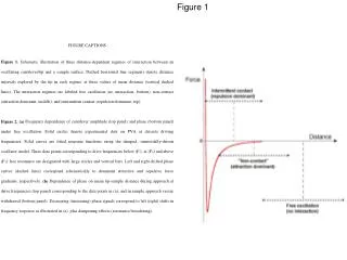
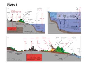
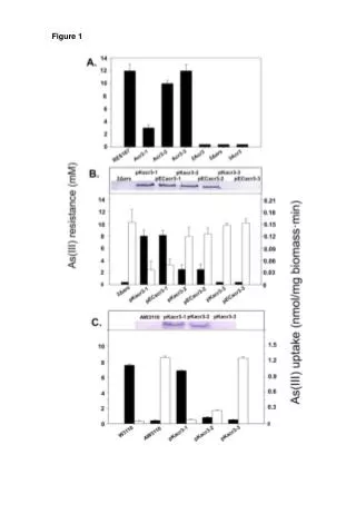
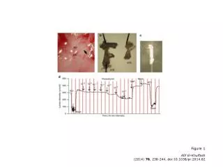
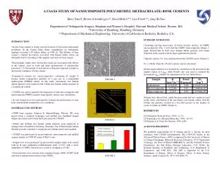
A USAXS STUDY OF NANOCOMPOSITE POLY(METHYL METHACRYLATE) BONE CEMENTSMary Turell, Heimo Schnablegger*, David Baker**, Lisa Pruitt**, Anuj BellareDepartment of Orthopaedic Surgery, Brigham and Women’s Hospital, Harvard Medical School, Boston, MA *University of Hamburg, Hamburg, Germany **Department of Mechanical Engineering, University of California at Berkeley, Berkeley, CA • SUMMARY OF RESULTS: • Scanning electron microscopy of freeze fracture surfaces of CMW1 microcomposite (Fig. 1; left) and the CMW1 nanocomposite (Figure 1; right) showed that in both cases barium sulfate particles were finely dispersed in the cement with no large agglomerates present. • Specific surface, Os, was calculated from the USAXS curves (Figure 2): • Os = p*K/Q where K = Porod constant, and Q = Invariant • Guinier approximation was invoked for calculation of the invariant in the angular region of 0-qmin while Porod’s law was used to estimate the invariant for qmax-infinity for calculation of Os (see Table below) • Fatigue tests showed that, under the processing and test conditions used in this study, substitution of the micrometer size barium sulfate with the 100nm size particles resulted in a two-fold increase in the number of cycles to failure in CMW1 (Figure 3). • REFERENCES • [1] Orthopedic Network News, 1999 , 11 (3). • [2] Topoleski et al, J Biomed Mater Res, 1990, 24:135 • [3] Demian et al, Trans Soc Biomat, 1995, p368 • ACKNOWLEDGEMENT • We gratefully acknowledge Dr. P. Jemian and Dr. J. Ilavsky for their assistance with USAXS measurements. The UNI-CAT facility at the Advanced Photon Source (APS) is supported by the University of Illinois at Urbana Champaign, Materials Research Laboratory (U.S. Department of Energy, the State of Illinois-IBHE-HECA, and the National Science Foundation), the Oak Ridge National Laboratory (U.S. DOE), the National Institute of Standards and Technology (U.S. Department of Commerce), and UOP LLC. The APS is supported by the U.S. Department of Energy, Office of Science, Office of Basic Energy Sciences, under Contract No. W-31-109-ENG-38. • INTRODUCTION • Acrylic bone cement is widely used for fixation of total joint replacement prostheses. In the United States alone, expenditures on orthopaedic implants exceeded 1.99 billion dollars in 1999 [1]. The lifetime of bone cement is limited by fracture associated with fatigue failure which can ultimately lead to loosening of the implant, and early revision surgery. • Fractographic studies have shown that cracks are associated with defects in the cement, such as voids and large agglomerates of radiopacifier particles [2, 3]. Prevention of such defects is therefore expected to improve the fracture toughness of bone cement. • Commercial cements are "microcomposites" containing 10 weight % barium sulfate radiopacifier particles of 1 mm size in a polymethyl methacrylate (PMMA) matrix. In this study, micrometer size barium sulfate particles were replaced with 100nm size barium sulfate particles in a commercial cement. • USAXS was used to quantify the dispersion of both microcomposite and nanocomposite PMMA cements using specific surface area calculations. • In vitro fatigue tests were performed to evaluate the performance of each bone cement under simulated in vivo conditions. • MATERIALS AND METHODS • CMW bone cements (Johnson & Johnson/Depuy, Warsaw, IN) were mixed using a standard techniques and molded into dumbbell shaped fatigue specimens and 0.5mm thick specimens for USAXS. • 100nm and 1000nm size barium sulfate particles were employed as radiopacifiers (Sachtleben, Duisberg, Germany) The 100nm barium sulfate ultrafine powder contained 2 weight percent sodium citrate anticoagulant. • USAXS was performed on microcomposite, nanocomposite and unfilled cement samples at UNICAT using 10 KeV x-rays. • Fatigue tests were performed on pre-notched samples under load control with an R ratio (minimum load/maximum load) of 0.03 with a stress amplitude of 15MPa. The load was cycled at a frequency of 2 Hz. • Freeze fracture cross sections were examined using a JEOL 6320FV field emission scanning electron microscope (SEM). FIGURE 1 FIGURE 2 FIGURE 3