Advancing Renal Research: Molecular Insights and Clinical Perspectives
Explore renal biopsy microdissection, transcriptomics, mRNA bioinformatics, RT-PCR, functional studies, and clinical insights in chronic kidney disease progression, apoptosis, inflammation, and hyperglycemia.

Advancing Renal Research: Molecular Insights and Clinical Perspectives
E N D
Presentation Transcript
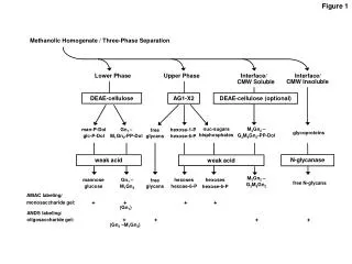
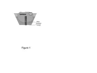
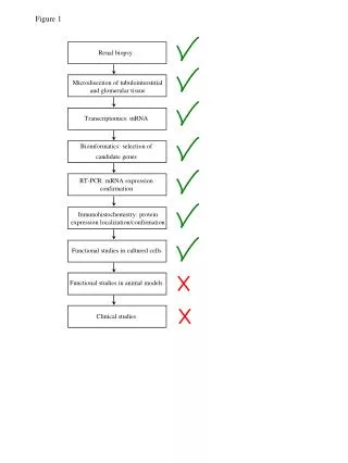
Renal biopsy Microdisection of tubulointerstitial and glomerular tissue Transcriptomics: mRNA Bioinformatics: selection of candidate genes RT-PCR: mRNA expression confirmation Inmunohistochemistry: protein expression localization/confirmation Functional studies in cultured cells Functional studies in animal models Clinical studies Figure 1
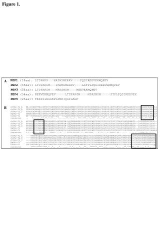
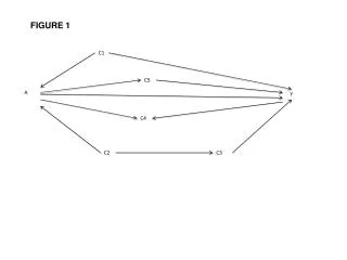
Osteoprotegerin TRAIL 14-3,3 eta CD74 GG2-1 Osteopontin Caspase 1 EIF4G2 TIA1 Clusterin STAT1 Tubulin-beta NRIF3 EMP3 APP DOCK1 Fas LOXL2 Osteoprotegerin MAPT Apolipoprotein E GADD45A PKC,zeta Sphingosine kinase 2 Prostaglandin E R-3 Cell death TRAIL NF-kB GG2-1 Figure 2 A) B) *
Figure 3 OPG Progression of chronic kidney disease Parenchymal renal cellapoptosis TRAIL Inflammation Hyperglycemia