Pyrabactin Mimicking Plant Hormone Interactions in Membrane Signaling Pathways
60 likes | 78 Vues
Investigation into the binding behaviors of PYL receptors and their key interactions with ABA, revealing insights into plant stress responses.

Pyrabactin Mimicking Plant Hormone Interactions in Membrane Signaling Pathways
E N D
Presentation Transcript
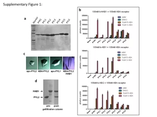
Supplementary Figure 1: b PYL5 Marker PYL4 PYL6 PYR1 PYL1 PYL2 PYL3 a None PYL2 PYL3 PYL4 PYL5 PYL6 PYR1 PYL1 c apo-PYL2 ABA-PYL2 apo-PYL1 ABA-PYL2 -HAB1 None PYL2 PYL3 PYL4 PYL5 PYL6 PYR1 PYL1 d HAB1 Mg2+ PYL2 pre- post- gelfiltration column None PYL2 PYL3 PYL4 PYL5 PYL6 PYR1 PYL1
Supplementary Figure 2: N N a C C gate gate latch latch b C N N C
Supplementary Figure 3: a K64 K64 N173 N173 E98 E98 E147 E147 b c latch + gate ABA-PYL2 apo-PYL2 ABA * ABA PYL2 G246D Mg2+ Mg2+ HAB1/PP2C HAB1/PP2C
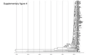
Supplementary Figure 4: a 680 nm 520-620 nm Singlet Oxygen b Streptavidin -Coated Donor Bead Ni-coated Acceptor Bead PYL2-H6 b-HAB1 IC50 = 250nM + untagged PYL2 competitor PYL2 680 nm Singlet Oxygen X no signal Streptavidin Ni-coated Acceptor Bead b-HAB1 PYL2 PYL2-H6
Supplementary Figure 5: ligand binding pocket gate relative luciferase activity latch
Supplementary Figure 6: a PYR1 residue number gate latch c b d