Download
1 / 33
400 likes | 892 Vues
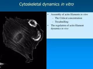
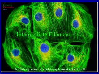
Regulation of Cytoskeletal Filaments. Pages 992-1010. Most cytoskeletal regulation is performed by accessory proteins that bind to either the filaments of their free subunits. Microtubule-organizing center (MTOC) – a specific intracellular location where microtubule nucleation occurs.

E N D
Regulation of Cytoskeletal Filaments Pages 992-1010
Most cytoskeletal regulation is performed by accessory proteins that bind to either the filaments of their free subunits. Microtubule-organizing center (MTOC) – a specific intracellular location where microtubule nucleation occurs
Nucleation of Microtubules by gamma-tubulin - end + end
The Centrosome, a MTOC 50 gamma-TuRC
Cross-section of a Centrosome The centrioles organize the centrosome matrix ensuring its duplication during each cell cycle Centrioles are composed of a short cylinder of modified microtubules and a large number of accessory proteins Neither fungi nor most plants have centrioles Centriole
Nucleation by the ARP Complex ARP – actin-related protein, each ARP is about 45% identical to actin
Binding of Profilin and Thymosin About 50% of actin in nonmuscle cells is in filaments and 50% as soluble monomers
Profin bound to Actin Monomer - Profilin binds to the opposite side of the ATP-binding site, blocking the side of the monomer that would associate with the filament minus end, allowing this complex to add onto a free plus end +
Effects of Stathmin on Microtubules Stathmin’s binding to tubulin is inhibited by the phosphorylation of stathmin
Organization of Microtubule Bundles MAP – Microtubule-associated protein MAPs have at least 1 domain that binds to the microtubule and another that projects outward
Localization of MAPS in a Neuron -MAP2 protein stained orange in the cell body and dendrites -tau stained green in the axon
Actin Filament Twisting Induced by Cofilin Cofilin – is a small protein that binds actin in a 1:1 ratio and destabilizes actin filaments Tropomyosin – an elongated protein that bind simultaneously to 7 actin monomers and stabilizes actin filaments
Filament Capping Changes Filament Dynamics CapZ – Capping protein Capping is regulated by intracellular signals, PIP2 (Phosphatidylinositol 4,5-bisphosphate) uncaps + ends
Proteins Binding Microtubule Ends Proteins that bind to the ends of microtubules can control microtubule positioning
Cross-linking Cyoskeletal Elements Red – Microtubules Blue – Intermediate Filaments Green – Cross-linking protein, Plectin Plectin also links IF to actin filaments and microtubules Filaggrin bundles keratin filaments in the epidermis of the skin to give it its toughness
Loss of Filamin Causes Abnormal Cell Motility Actin formed by filamin is required for cells to extend the thin sheet-like membrane projections call lamellipodia
Microtubule Severing Red - Microtubules
Actin Filament Severing by Gelsolin -Activated by high levels of cytoplasmic calcium -No energy needed -Gelsolin is removed by PIP2 Severing of microtubules by Katanin -made up of 2 subunits, one for severing and the other for targeting it -the process requires ATP
Focal Contacts in Fibroblasts Focal contacts –highly specialized type of attachment between actin filaments and the extracellular matrix