The Development and Impact of Cancer Cells in the Brain: A Visual Overview
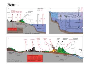
This visual guide illustrates the interplay between neurons, astrocytes, oligodendrocytes, and cancer cells in the brain. Starting with the structure of healthy brain cells and their supportive roles, the images transition to showcase the transformation of astrocytes and the process of cancer cell mitosis. As cancer cells proliferate and create tumor masses, they outpace their blood supply, starving surrounding healthy cells. This sequence culminates in the necrosis of normal brain tissue, emphasizing the aggressive nature of tumors and their far-reaching impact on brain health.

The Development and Impact of Cancer Cells in the Brain: A Visual Overview
E N D
Presentation Transcript
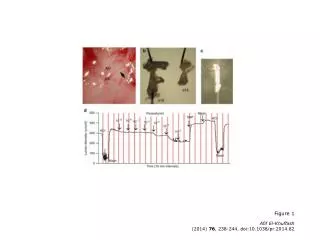
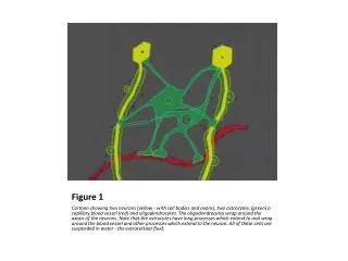
Figure 1 Cartoon showing two neurons (yellow - with cell bodies and axons), two astrocytes, (green) a capillary blood vessel (red) and oligodendrocytes. The oligodendrocytes wrap around the axons of the neurons. Note that the astrocytes have long processes which extend to and wrap around the blood vessel and other processes which extend to the neuron. All of these cells are suspended in water - the extracellular fluid.
Figure 2 Transformed astrocyte pulls in its processes and detaches itself from neuron and blood vessel, in order to multiply.
Figure 3 Cancer cell undergoes mitosis where one cell becomes 2 cells. It is the reproductive method of all single celled animals and all cells within organ systems of every species in the animal kingdom.
Figure 4 Two cancer cells following mitosis. Each are completely independent and can survive without the assistance of each other and the normal cells in their environment.
Figure 5 Cancer cells are capable of movement as well as mitosis but probably not at the same time. They have to stop moving in order to reproduce.
Figure 6 Mitoses have produced more cancer cells, creating the beginning stages of a brain tumor. Cell movement has allowed cancer cells to spread to new (and less polluted) areas.
Figure 7 Mitotically active cancer cells have not travelled away but have stayed in the same location now require more nourishment than available only through the extracellular fluid. Cytokines (such as the so-called Tumor Angiogenesis Factor) are produced by the tumor cells and probably normal cells also. This results in mitosis (reproduction) of the blood vessel endothelial cells which form new blood vessels to supply the tumor.
Figure 8 A solid mass of tumor cells lumped together, growing outward as more cells are added to the mass by mitosis. Newly formed blood vessels supply the mass with everything the cells need to survive and grow. The nerve cells, astrocytes and oligodengroglial cells are now being starved and will die.
Figure9 The tumor mass has killed the background neurons and other normal cells. It has outpaced its blood supply and the center of the tumor has undergone necrosis. This picture does not show the fact that this tumor mass is surrounded by isolated tumor cells which can extend a great distance (up to 3 inches - 7 cms.) into the surrounding functional brain tissue.
Info From: • http://www.brainlife.org/fulltext/1995/kelly_gliomas.htm