Suppl Figure 1
Suppl Figure 1. Growth characteristics of CHO-K1 wt and CKiD cells:

Suppl Figure 1
E N D
Presentation Transcript
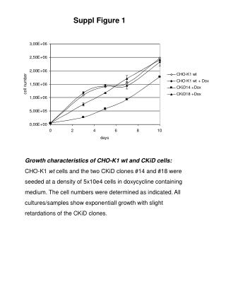
Suppl Figure 1 Growth characteristics of CHO-K1 wt and CKiD cells: CHO-K1 wt cells and the two CKiD clones #14 and #18 were seeded at a density of 5x10e4 cells in doxycycline containing medium. The cell numbers were determined as indicated. All cultures/samples show exponentiall growth with slight retardations of the CKiD clones.
Suppl Fig. 2 Antibody production [µg/ml] Small scale protein production For generation of antibody producing cells, clone #18 was stably transfected with pAkNKis and cultivated +/- Dox according to the procedure specified in the text. To assess production, the cells were seeded with a concentration of 105 cells in a 12-well dish and cultivated at confluency for 7 days in presence and absence of Dox. The concentration of produced antibody was determined from triplicates and is given in µg/ml.
Suppl Figure 3 IgG standard 0,1µg clone #18 -dox clone #18 +dox CHO-K1 wt heavy chain light chain Antibody expression: Antibody expression from clone #18 cells transfected with pAkNKis and selected in presence and absence of Dox was determined by Western Blot analysis. For this purpose, the supernatants of clone #18 derived cell populations cultivated in presence and absence of Dox and of CHO-K1 cells (negative control) were seperated on an SDS-PAGE and tranferred to a membrane. An IgG standard was used as a positive control. For detection, Goat-Anti-Human IgG (H+L) HRPO labelled antibody (CALTAGTM Laboratories, Code No. H10307) was employed and visualized using Quantity OneR system (Biorad). Note that the production of the +Dox samples was confirmed upon longer exposition (data not shown).