Restoration of Wound-Induced MAP Kinase Activation in Complemented ap2c1 Plants
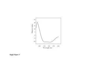
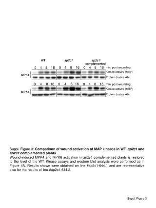
This study investigates the wound activation of MAP kinases MPK4 and MPK6 in wild-type (WT), ap2c1, and complemented ap2c1 plants. Our findings reveal that the wound-induced activation of MPK4 and MPK6 in complemented ap2c1 plants is restored to WT levels. Kinase assays and western blot analyses corroborate this restoration, demonstrating the efficacy of the complementation. Results were derived from line #ap2c1-644.1, which are also representative of results from line #ap2c1-644.2.

Restoration of Wound-Induced MAP Kinase Activation in Complemented ap2c1 Plants
E N D
Presentation Transcript
WT ap2c1ap2c1/ complemented 4 4 4 4 8 8 8 8 16 16 16 16 0 0 0 0 min. post wounding Kinase activity (MBP) MPK4 4 8 16 0 Protein (native Ab) 4 8 16 0 min. post wounding Kinase activity (MBP) MPK6 Protein (native Ab) Suppl. Figure 3: Comparison of wound activation of MAP kinases inWT, ap2c1 and ap2c1 complemented plants Wound-induced MPK4 and MPK6 activation in ap2c1 complemented plants is restored to the level of the WT. Kinase assays and western blot analysis were performed as in Figure 4A. Results shown were obtained on line #ap2c1-644.1 and are representative also for the results of line #ap2c1-644.2. Suppl. Figure 3