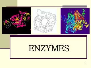
Deconstruction by Enzymes 1: Cellulase
1.07k likes | 2.2k Vues
Deconstruction by Enzymes 1: Cellulase. 2007 Oct. 19 Speaker: Dr. Po-Huang Liang. CONTENT. INTRODUCTION FUNDAMENTALS -Structure and Composition of Cellulosic Biomass -Cellulolytic Organisms -Cellulase Enzyme Systemes -Regulation of Cellulase Production APPLICATION OF CELLULASES

Deconstruction by Enzymes 1: Cellulase
E N D
Presentation Transcript
Deconstruction by Enzymes 1:Cellulase 2007 Oct. 19 Speaker: Dr. Po-Huang Liang
CONTENT INTRODUCTION FUNDAMENTALS -Structure and Composition of Cellulosic Biomass -Cellulolytic Organisms -Cellulase Enzyme Systemes -Regulation of Cellulase Production APPLICATION OF CELLULASES -Recombinant Cellulolytic Strategy -Methodology for Studying Cellulase Properties CLOSING COMMENT
INTRODUCTION Need for Alternative Energy Source With the hike in oil price around the world in the 1970s and the realization that the world’s oil supply is finite, the quest for alternative fuels began in 1975. The amount of solar energy received at the earth’s surface far exceeds the amount of present human usage 2.5x10²¹ Btu/year >> 2.0x10¹⁷ Btu/year The amount of energy from sun is stored as carbon via photosynthesis, which results in production of plant biomass having cellulose as the major component
Cellulase Strategy of converting cellulose into fuel Biomass Biofuel Glucose
Structure and Composition of Cellulosic Biomass In plants, the cell wall is constructed primarily from a carbohydrate polymer called cellulose, and the cell wall can therefore also function as a carbohydrate store for the cell. genomics.energy.gov/gallery/gtl/originals/420.jpg
Plant cell wall structure • Plants form two types of cell wall that differ in function and in composition: • - primary walls surround growing • and dividing plant cells. • secondary wall is much thicker • and stronger than primary wall and • accounts for most of the • carbohydrate in biomass. • Middle lamella is a specialize region associated with the cell walls and is • shared by neighboring cells. • Plasmodesmata is the small passages penetrate the middle larmella as well ass the primary and secondary cell walls, providing pathways for transporting cytoplasmic molecules from one cell • to another. tainano.com/.../image036.gif
Plant cell wall structure middle lamella primary cell wall pectin cross-linking glycan plasma membrane cellulose microfibrils Primary Cell Walls The main chemical components of the primary plant cell wall include cellulose. In addition, the cell wall contains two groups of branched polysaccharides, the pectins and cross-linking glycans. Organized into a network with the cellulose microfibrils, the cross-linking glycans increase the tensile strength of the cellulose, whereas the coextensive network of pectins provides the cell wall with the ability to resist compression. The middle lamella is rich in pectins. micro.magnet.fsu.edu/cells/plamts/cellwall.html
Plant cell wall structure middle lamella primary cell wall hemicellulose protein plasma membrane lignin Secondary Cell Wall The secondary cell walls contain cellulose, hemicellulose and pectin as well as the primary cell walls, albeit in different proportion. The cellulose fibrils are embedded in a network of hemicellulose and lignin to make a further strengthened form. Numerous technical challenges must be overcome to enable the efficient utilization of secondary walls for energy production. cellulose www.ccrc.uga.edu/~mao/intro/ouline.htm
Structure of cellulose Cellobiose Glucose Elementary cellulose fibril Lignin Hemicellulose 7-30 nm Microfibril Cellulose is a linear condensation polymer consisting of glucose subunit linked by b-1, 4-glycosidic bonds. Coupling of adjacent cellulose molecules by the hydrogen bonds and van der Waal’s forces results in a parallel alignment and a crystalline structure. Approximately 30 individual crystalline cellulose moleculesare packed into larger units called microfibrils, which are in turn assembled into the familiar cellulose fibers.
Structure of cellulose Amorphous region Crystalline domain Crystalline domain The microfibril chain are oriented in parallel and form highly ordered, crystalline domains interspersed by more disordered, amorphous regions. The native, crystalline form of cellulose has a structure designated as type I, which can be converted to other crystalline forms (II-IV) by various treatments. Cellulose I can be transformed into cellulose II by alkali treatment and into cellulose III by supercritical ammonia treatment. Cellulose IV could be prepared by heat treatment in glycerol after transformation into cellulose II or cellulose III but cannot be transformed directly from cellulose I. Native cellulose has two distinct crystallite form, Ia and Ib, which differ in their intermolecular hydrogen bonding pattern. Form Ia is dominant in bacterial and algal cellulose and form Ib is dominant in higher plants.
Microorganism - Bacteria: fermentative anaerobes (Clostridium, Ruminococcus, Fibrobacter) aerobic gram-positive (Cellulomonas, Thermobifida) aerobic gliding (Cytophaga, Sporocytophaga) Clostridium Ruminococcus Cellulomonas Cytophaga Cellulolytic Organisms In plants, cellulases hydrolyze their cell walls at various developmental stages (e.g., bean abscission, fruit ripening and abscission, and pedicel abscission). For microorganisms and animals, hydrolysis and utilization of cellulose by cellulsas can provide usable energy to them.
Microorganism - Protozoa: ciliate (Diplodinium, Eudiplodinium) Protozoa of Diplodinium and Eidinium type attached to fodder molecules in rumen liquid (Dobicki et al., 2006) Piromyces Anaeromyces Chaetomium Phanerochaete Trichoderma - Fungi: monocentric (Neocllimastix, Piromyces, Caecomyces) policentric (Orpimomyces, Anaeromyces) Ascomycetes (Bulgaria, Chaetomium, Helotium) Basidiomycetes (Coriolus, Phanerochaete, Serpula) Deuteromyces (Aspergillus, Cladosporium, Penicillium, Trichoderma)
Nasutitermes Cryptocercus Ergates Animal - Arthropods: termite (Coptotermes, Nasutitermes, Neotermes, Reticulitermes) cockroach (Cryptocercus, Panesthia) beetle (Ergates) Fungus (Termitomyces) Termite nest (Odontotermes) (Ohkuma, 2003) Symbioses - higher termite-bacteria - lower termite-bacteria-protozoa - lower termite-fungus
Animal - Molluscs snail (Achatina, Helix, Levantina) bivalve (Mytilus,Xylophaga) sea slug (Dolabella) marine periwinkle (Littorina)
General Feature Microorganisms - cellulase systems including a multiplicity of enzyme components - cellulase systems exhibiting higher collective activity than the sum of the activities of individual enzymes - most cellulases containing both catalytic and carbohydrate- binding modules - cellulases often associated with each other and with the surface of cellulolytic microorganisms Termite - endogeneous cellulase excreted from the salivary glands or the mid-gut - cellulases of termite origin belonging to glycosyl hydrolase family 9 - cellulases containing a single catalytic domain and lacking the ancillary domains such as cellulose-binding domain - cellulase systems consisting of endogeneous cellulases from termite and cellulolytic enzymes of gut protists Cellulase Enzyme Systems Since cellulose cannot get into the cells, cellulolytic enzymes are by necessity secreted into the medium or bound to the outside surface of cellulolytic organisms.
Components of cellulase systems Cellulases are distinguished from other glycoside hydrolases by their ability to hydrolyze b-1, 4-glucosidic bind between glucosyl residues. Based on mode of catalytic action and on structural properties, three major types of enzymatic activities are found: (i) Endo-b-1, 4-glucanase (E.C. 3.2.1.4) cleaves at internal amorphous site in the cellulose polysaccharide chain, generating oligosaccharides of various lengths and consequently new chain end (ii) Exoglucanase (cellodextrinases E.C. 3.2.1.74 or cellobiohydrolase E.C. 3.2.1.91) acts in a processive manner on the reducing or nonreducing ends of cellulose polysaccharide chain, releasing either glucose or cellulobiose (iii) b-Glucosidase (E.C. 3.2.1.21) hydrolyzes soluble cellodextrins and cellobiose to glucose
Components of cellulase systems Reaction mechanisms It is generally assumed that the hydrolysis reaction catalyzed by cellulase proceeding via an acid-base mechanism involving two residues, one as a general acid catalyst and another as a nucleophile. Glu-555 Glu-555 Asp-201 Asp-201 Catalytic mechanism of C. thermocellum endoglucanase CelD as a paradigm of b-glucanase acting (Beguin and Aubert, 1992))
Microorganism cellulase system Microorganism have adapted different approaches to effectively hydrolyze cellulose. There are two different systems: Noncomplexed cellulase systems The microorganisms, such as cellulolytic filamentous fungi and actinomycete bacteria, have the ability to penetrate cellulolytic substrate and produce ”free” cellulases, with or without cellulose- binding modules. Crystalline Amorphous Crystalline Endoglucanase Nonreducimg end b-glucosidase Exoglucanase Reducing end Glucose Cellobiose Exoglucanase Cello-oligosaccharides (Lynd et al., 2002)
Microorganism cellulase system Complexed cellulase systems The microrganisms lack the ability to effectively penetrate cellulosic material and perhaps exists under a condition in the present of competition from other microorganisms and with limited ATP available for cellulase synthesis. This could have led to the development of “complexed” cellulase systems (called “cellulosome”). Bacterium cell wall Scaffodin Endoglucanase (with dockerin) Cellobiose/cellodextrin phosphorylase Exoglucanase (with dockerin) Cohesin moiety Glucose Cellobiose Exoglucanase (with dockerin) Cello-oligosaccharides Carbohydrate-binding module (CBM) Crystalline Amorphous Crystalline (Lynd et al., 2002)
Microorganism cellulase system Cellulose-binding domain (CBD) CBDs provide a specific means for linking enzymes or other proteins on cellulose. These domains are usually located at the NH2 or COOH terminus of the enzymes and are often separated from the catalytic domains by glycosylated, Pro/Thr/Ser-rich linker segments. Hydrogen bond formation and van Waals interactions are the main driving forces for binding. In proteins that possess hydrolytic activity the CBD concentrates its catalytic domains on the surface of the insoluble cellulose substrate. In proteins that have no hydrolytic activity, CBD is part of a scaffolding subunit that organizes the catalytic subunits into a cohesive multienzyme complex know as a cellulosome. Based on amino acid sequences, binding specificity and structures, CBD can be divided to 16 different families among 48 different carbohydrate-binding modules.
Microorganism cellulase system Cellulose-binding domain (CBD) Most of the CBDs found belong to the four major families: (Shoseyov and Warren, 1997)
Microorganism cellulase system Cellulosome The plant cell wall degrading enzymes in most anaerobic microorganisms associate into a supramolecular complex, termed the “cellulosome” with a molecular mass higher than 2 MDa. The cellulosomes are associated with the cell surface and mediate cell attachment to the insoluble substrate and degrade it to soluble products which are then absorbed. cellulosomes Cell membrane S-layer Cell anchoring proteins cellulosomes cellulose (Bayer et al., 1998)
Microorganism cellulase system Cellulosome The principal component of the cellulosome is a scaffoldin subunit that contains cohesin modules and also frequently includes a carbohydrate-binding module. The enzymatic subunits of the cellulosome contain a complementary type of module, the dockerin domain, that is responsible for attachment to the cohesin modules of scaffoldin. N N C C bacterial cell Type I Dockerin Type I Cohesin X Module Scaffoldin SLH Module Type II Dockerin Type II Cohesin Cellulose-binding domain Catalytic domain (Demain et al., 2005)
Microorganism cellulase system Cellulosome Recently, several different modular structures of scaffoldin have been described in various anaerobic microognisms. Clostridium thermocellum CipA C. celluloticum CipC 1 2 3 4 5 6 7 8 9 1 2 3 4 5 6 7 8 X II X X CBD CBD Acetivibrio cellulolyticus CipV C. josui CipJ 1 2 3 4 5 6 X 1 2 3 4 5 6 7 X II GH9 CBD CBD 1 2 3 4 5 6 7 8 9 10 11 II Bacteroides cellulosolvens ScaA C. cellulovorans CbpA 1 2 3 4 5 6 7 8 9 X X X CBD X CBD
Microorganism cellulase system Cellulosome The cellulosome of most anaerobic bacteria is in essence a cell- surface component which need a cell-surface protein to mediate the its binding. The most studied specie, Clostridium thermocellum, has been identified four cell-surface proteins and one scaffoldin that are relative to this binding. OlpB Orf2p OlpA SdbA Bacterial Cell (Demain et al., 2005) (Bayer et al., 1998)
Termite cellulase system The presence of gut protists is important to termite survival on a diet of cellulose as their energy and carbon source. Termites grind and crunch their ingested material, which may enhance digestion by increasing the amount of surface that can be accessed by cellulolytic enzymes. Probably, the ingested cellulose can be partially degraded by the endoglucanase of termite origin, and the cellulose not hydrolyzed in the anterior portion of the gut then travels to the hindgut, where it can be endocytosed and fermented by the symbiotic microrganisms. Hindgut Protist Mid-gut Cellulose decomposition Ingested cellulose Gring Crunch Cellulolytic material endogeneous cellulase CO2 H2 Acetate CH4 Absorbed by termite
Carbon Source Regulation Cellulose and Derived Metabolites For most microorganisms, cellulase synthesis generally requires the presence of cellulose or its soluble metabolites. Cellobiose functions as an inducer is more complex because at high level it inhibits cellulase production. In case of fungi, cellulose induces cellulase synthesis in germinating conidia, but not in mycelium. Sophorose (b-1,2-glucobiose), that is formed via the transglycosylation of cellobiose by a b-glucosidase, was identified as a strong inducer of cellulase formation in fungi. Easily Metabolized Substrates In most moicroorganisms, cellulase synthesis is blocked in presence of soluble substrate such as glucose. Regulation of Cellulase Production
Intracellular Molecules Transcriptional Factors ACEI and ACEII were identified the ability to bind to the promoter region of fungal cellulase gene thus can stimulate the expression of cellulase gene. Gene Cluster In the case of anaerobic bacteria, several gene clusters have been found, suggesting the existence of operons as units of gene regulation. Negative control For clostridia, three protein, GlyR1, GlyR2, and GlyR3 were identified as regulatory proteins containing two major domains, a sugar-binding domain and a DNA-binding domain. These proteins inhibit the cellulase production by binding to the promoter region of cellulase gene.
Intracellular Molecules Carbon Catabolite Repressor Cre1 or CreA was identified as an inhibitor for the transcription of cellulase gene of fungus. ATP and cAMP In the case of fungi, extracellular cellulase was repressed at intracellular ATP concentration at a high level and cAMP played a role in derepression of enzyme synthesis. Enzyme Inhibitor Nojirimycin and Gluconolactone The inhibition of b-glucosidase activity by these components may prevent induction by celloluse.
Cellulases Some cellulase enzymes may play a role in the formation of the inducer for other enzymes. In T. reesei, Fowler and Brown (1992) suggested that BGL1 may be partially responsible for formation of the inducer because the deletion of bgl1 gene resulted in decreased endoglucanase activities. Later, Seiboth et al. (1997) revealed that deletion of cbh2 and eg2 genes prevented the expression of other cellulase genes. In R. flavefaciens, Doerner et al. (1992) reported that the celA and celC genes were expressed constitutively while expression of the celB and celD was induced by cellulose. In C. thermocellum, Mishra (1991) revealed that transcription of several cel genes was induced sequentially when cellobiose concentration in the medium became limiting. Transcription of celA started first, followed by celD and celF, and finally celC.
Summery Cellulose first undergoes limited hydrolysis by cellulases constitutively produced in low amounts. The soluble hydrolysis products thus generate and cause induction of cellulase synthesis. Catabolite repression of cellulase genes occurs in the presence of glucose and may be regulated by cAMP.
APPLICATION OF CELLULASES
Substrate for cellulase activity assays Substrate for cellulase activity assays can be divided into 2 categories, based on their solubility in water. Methodology for Studying Cellulase Properties a RS, reducing sugars; TSS, total soluble sugars. (Zhang et al., 2006)
Substrate for cellulase activity assays a RS, reducing sugars; TSS, total soluble sugars. (Zhang et al., 2006)
Cellulase activity assays All existing cellulase activity assays can be divided into three types: 1) the accumulation of products after hydrolysis - reducing sugars - total sugars - glucose 2) the reduction in substrate quantity - gravimetry - chemical methods 3) the change in the physical properties of substrates - swollen factor - fiber strength - structure collapse - turbidity - viscosity
Cellulase activity assays – accumulation of hydrolysis products Reducing sugars depend on the reduction of inorganic oxidants such as cupric ions (Cu2+) or ferricyanide, which accepts electrons from the donating aldehyde groups of reducing cellulose chain ends. The common colorimetric reducing sugar assays G: reducing sugar; DNS: dinitrosalicyclic acid; PAHBAH: 4-hydroxybenzoylhydrazine; BCA: 2,2’-bicinchroninate
Cellulase activity assays – accumulation of hydrolysis products Total soluble sugars Phenol or anthrone in the presence of sulfuric acid can be used for the quantitative colorimetric microdetermination of sugars and their methylderivatives, oligosaccharides, and polysaccharides, to give an orange-yellow or blue-green color. The common colorimetric total sugar assays G: reducing sugar
Cellulase activity assays – accumulation of hydrolysis products Glucose Assay Enzymatic Glucose Assays depend on the glucose oxidase-peroxide reaction for the determination of glucose concentrations byusing coupled hexokinase (HK) and glucose-6-phosphate dehydrogenase(PGHD). The colorimetric enzymatic glucose assays G: reducing sugar HPLC After post-hydrolysis conversion to glucose
Cellulase activity assays – loss of substrate Gravimetry uses precipitation or volatilization method based on the determination of a substance of known composition that is chemically related to the sugar. The standard deviation of this method is strongly associated with sample weight. Chemical Method includes the phenol-H2SO4 and the anthrone-H2SO4 method for residual cellulose, and HPLC quantitative saccharification for different carbohydrate components.
Cellulase activity assays – physical cellulose properties Swollen Factor measures by alkali uptake. Structure Collapse measures the reduction in tensile strength of cellulotic fiber. Turbidity measures a reduction in the absorbance of particle suspension during the hydrolysis process. Amorphous cellulose is recommended for this assay. Viscosity measures a reduction in substrate viscosity. Soluble cellulose derivatives are recommended for this assay.
Endoglucanase activity assays Endoglucanases cleave intramolecularb-1,4-glucosidic linkages randomly, and their activities can be measured based on a reduction in substrate viscosity and/or an increase in reducing ends determined by a reducing sugar assay. Because exoglucanases also increase the number of reducing ends, it is strongly recommended that endoglucanase activities be measured by both methods. CMC, a soluble high DP (degree of polyerization) cellulose derivative, is often recommended as a good substrate for endoglucanase activities. Soluble oligosaccharides and their chromophore-substituted substrates, such as p-nitrophenyl glucosides and methylumbelliferyl-b-D-glucosides, are also to measure endoglucanase activities based on the release of chromophores or the formation of shorter oligosaccharide fragments, which are measured by HPLC or TLC. Endoglucanase activities can also be easily detected on agar plates by staining residual polysaccharides (CMC, cellulose) with various dyes, such as Congo red, because these dyes are adsorbed only by long chains of polysaccharides.
Exoglucanase activity assays Exoglucanases cleave the accessible ends of cellulose molecules to liberate glucose and cellobiose. During chromatographic fractionation of cellulase mitures, enzymes with little activity on soluble CMC, but showing relatively high activity on Avicel, are usually identified as exoglucanase. There is no substrates specific for exoglucanase within the cellulase mixtures. Soluble oligosaccharides and their chromophore-substituted substrates, such asp-nitrophenyl-b-D-cellobioside,4-methylumbelliferyl-b-D-lactoside, 4-methylumbelliferyl-b-D-aglyconesand 4-methylumbelliferyl-b-D-glycosides, are also to measure exoglucanase activities, which can be differentiated from endoglucanase activities by the aid of their specific inhibitor such as cellobiose. But different exoglucanases have different activities on these substrates.
b-glucosidase activity assays b-glucosidase hydrolyze soluble cellobiose and other cellodextrins in the aqueous phase. These enzymes are very amenable to a wide range of simple sensitive assay methods, based on colored or fluorescent products released from p-nitrophenyl b-D-1,4-glucopyranoside, b-naphthyl-b-D-glucopyranoside, 6-bromo-2-naphthyl-b-D-glucopyranoside, and 4-methylumbelliferyl-b-D- glucopyranoside. Cellobiose, which is not hydrolyzed by endoglucanases and exoglucanases, is also used as a substrate for b-glucosidase activity assays by determining the increase of reducing sugar.
Total cellulase activity assays The total cellulase activity assays are always measured using insoluble substrates, including pure cellulosic substrates such as Whatman No. 1 filter paper, cotton fiber, microcrystalline cellulose, bacterial cellulose, algal cellulose; and cellulose-containing substrates such as dyed cellulose, a-cellulose, and pretreated lignocellulose. The most common total cellulase activity assay is filter paper assay (FPA) which requires a fixed amount (2mg) of glucose released from a 50-mg sample of Whatman No.1 filter paper. a-cellulose and pretreated lignocellulose are often used to evaluate the digestibility of a reconstituted cellulase mixture for a prolonged reaction. Dyed celluloses and fluorescent-dyed celluloses are widely used for determining sugar inhibition for total cellulase.
Applied objective: CBP-compatible strains of use for industrial processes Strain characterization and improvemetn Engineered strains able to utilize cellulose and produce a desired product at high yield Native strategy: Metabolic engineering to improve product yields, titer ect. Recombinant strategy: Heterologous cellulase expression Fundamentals of microbial cellulose utilization Microbes with good substrate utilization properties: Cellulase production, utilization of hydrolysis products (e.g. thermophiles) Microbes with good product-producing properties: High product yields, titers ect. (e.g. yeasts) Underlying fundamental issue: Understand cellulose hydrolysis at a microbial rather than an enzymatic level Recombinant Cellulolytic Strategy (Lynd et al., 2005)
Strain-donor The most studied cellulolytic organismsand their properties are listed as follow:
Strain-host The most studied host microorganisms and their functions are listed as follow:
Cellulase improvement Two methods are available for improving the properties of individual cellulase components:1) rational design and 2) directed evolution. Rational design is the eariliest approach to protein engineering and requires detailed knowledge of protein structure. (Zhang et al., 2006)
Cellulase improvement Directed evolution is independent of knowledge of enzyme structure and of interactions between enzyme and substrate, and is developing tools to correctly evaluate the performance of mutants generated by recombinant DNA techniques. (Zhang et al., 2006)