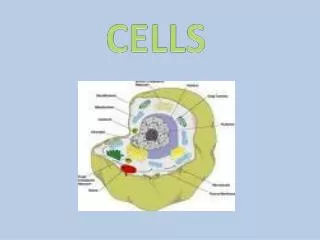
Overview of Animal Cells: Structure, Function, and Organelles
Animal cells are fundamental units of life, with an estimated 75 trillion cells in the human body, each varying in size and function. Key components include the cell membrane, which regulates substance movement; cytoplasm, serving as the site of metabolic processes; and vital organelles like the nucleus, endoplasmic reticulum, ribosomes, mitochondria, and lysosomes. This overview covers the essential structures and their respective functions in maintaining cell integrity and supporting cellular activities.

Overview of Animal Cells: Structure, Function, and Organelles
E N D
Presentation Transcript
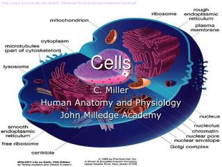
http://www.amersol.edu.pe/class09/_09sschee/7th/science/cell/images/animalcell.gifhttp://www.amersol.edu.pe/class09/_09sschee/7th/science/cell/images/animalcell.gif Cells C. Miller Human Anatomy and Physiology John Milledge Academy
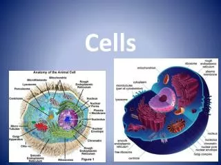
Introduction • 75 trillion cells in the body • Vary in size, shape, content, and function • Most common structures: nucleus and cytoplasm http://www.crossroadsinitiative.com/pics/Theology_of_the_Body_Christopher_West_DaVinci.jpg
I. The Cell Membrane • Function: maintains “wholeness”; controls entrance and exit of substances 1. Semipermeable or selectively permeable http://library.thinkquest.org/C004535/media/cell_membrane.gif
I. Cell Membrane B. Structure 1. lipids and proteins 2. Phospholipid bilayer a. hydrophilic phosphate heads b. hydrophobic fatty acid tails 3. Has an oily characteristic a. lipid soluble molecules, but not water soluble can freely pass b. embedded cholesterol
I. Cell Membrane B. Structure 4. Proteins a. fibrous b. carrier c.glycoproteins http://lhs.lps.org/staff/sputnam/Biology/U3Cell/membrane_1.png
II. Cytoplasm • jelly-like fluid fills the cell • Site of metabolic activities http://sciencecity.oupchina.com.hk/biology/student/glossary/img/cytoplasm.jpg http://sun.menloschool.org/~cweaver/cells/c/cytoplasm/jrcytoplasm.jpg
III. OrganellesA. ENDOPLASMIC RETICULUM • Structure: membrane-bound sacs/canals • Function: • Rough: ribosomes site of protein synthesis b. Smooth: lipid synthesis http://www.fairfield.k12.ct.us/tomlinson/ctomlinson03/CellProject04/Per2/2MS/endoplasmic_reticulum.jpg http://www.lifesci.sussex.ac.uk/home/Julian_Thorpe/tem3.jpg
III. OrganellesB. RIBOSOMES • Structure: • May be attached or free floating particles • made of proteins and RNA • Function: protein synthesis http://iupucbio2.iupui.edu/anatomy/images/Chapt02/FG02_11.jpg http://www.rockefeller.edu/rucal/journey/ribosomes.gif
III. OrganellesC. GOLGI APPARATUS http://www.fairfield.k12.ct.us/tomlinson/ctomlinson03/CellProject04/Per2/2JD/golgi.jpg • Structure: stack of flattened membranes • Function: package and refine proteins • Transport vesicles move refined proteins to other cell parts http://biology.unm.edu/ccouncil/Biology_124/Images/golgiapparatusmicroscope.jpeg
III. OrganellesD. MITOCHONDRIA • Structure: fluid-filled elongated sac • Double membrane - cristae: folded layer • Function: ENERGY!! http://www.abcbodybuilding.com/magazine03/mitochondria.jpg http://cellbio.utmb.edu/cellbio/mitmor4.jpg
III. OrganellesE. LYSOSOMES • Structure: tiny membranous sac • Function: enzymes break down foreign molecules and wastes http://www.visualsunlimited.com/images/watermarked/350/350789.jpg http://www.fairfield.k12.ct.us/tomlinson/ctomlinson03/CellProject04/Per4/4BRL/lysosomes.jpg
III. OrganellesF. CENTROSOME • Structure: near nucleus, 2 hollow cylinders form centrioles • Function: role in cellular reproduction http://www-ijpb.versailles.inra.fr/en/sgap/equipes/cyto/images/centrosome.jpg
III. OrganellesG. VESICLES http://www-cse.stanford.edu/classes/sophomore-college/projects-01/cellular-automata/beginning/vesicles.jpg • Structure: membranous sac formed by cell membrane • Function: move materials throughout cell http://www.visualsunlimited.com/images/watermarked/284/284900.jpg
III. OrganellesH. Other cellular structures • Cilia • Structure: short, hair-like projections w/ “to and fro” movements • Function: movement; Ex: epithelial cells http://www.uib.no/med/mic/gallery/pictures/SEM/SEM-large/ciliated-epithelium_870.jpg
III. OrganellesH. Other cellular structures 2. Flagella • Structure: long, whiplike projection w/ wave-like movements • Function: movement; Ex: sperm cells http://images.livescience.com/images/050330_sperm_egg_01.jpg http://newsimg.bbc.co.uk/olmedia/400000/images/_404096_single_sperm300.jpg
III. OrganellesH. Other cellular structures 3. Microfilaments • Structure: tiny protein rods • Function: cellular movement 4. Microtubules (spindle fibers) • Structure: thick tubes of protein • Function: maintain cell structure; strength to motile processes These make up the CYTOSKELETON!
http://www.cdli.ca/~dpower/cell/nucleus.jpg IV. The Nucleus • Structure: large, spherical; w/in nuclear envelope • porous • Nucleolus: dense area of ribosome synthesis; ”little nucleus” • Chromatin→ Chromosomes http://blogofthe.s57.xrea.com/media/1/20040407-nucleus.jpg
V. Cell Cycle A. Interphase • G1: Growth • S: Synthesis of DNA • G2: Growth and mitosis prep • M: Mitosis (nuclear division) • C: Cytokinesis (cytoplasm division) http://www.isd2184.net/~jensenje/biology/BioReviewWeb/U2ReviewWeb/mitosisWeb/Images/Cellcyclediagram.jpg
Prophase Metaphase 3. Anaphase 4. Telephase V. Cell CycleB. MITOSIS http://www.houghtonmifflinbooks.com/booksellers/press_release/studentscience/gif/mitosis1.gif
PROPHASE INTERPHASE METAPHASE MITOSIS ANAPHASE TELOPHASE
V. Cell CycleC. CYTOKINESIS • Two identical daughter cells formed. http://raven.zoology.washington.edu/celldynamics/research/cytokinesis/images/SdTeloAT.jpg
http://training.seer.cancer.gov/ss_module08_lymph_leuk/images/illu_blood_cell_lineage.jpghttp://training.seer.cancer.gov/ss_module08_lymph_leuk/images/illu_blood_cell_lineage.jpg D. DIFFERENTIATION http://training.seer.cancer.gov/ss_module08_lymph_leuk/images/illu_blood_cell_lineage.jpg
V. Cellular MovementsA. PASSIVE TRANSPORT • No cellular energy (ATP) required. • Diffusion: molecules move from areas of high concentration to low (down the concentration gradient) • Equilibrium: solute evenly distributed w/in solvent • Ex: exchange of O2/CO2 in the lungs
DIFFUSION http://www.indiana.edu/~phys215/lecture/lecnotes/lecgraphics/diffusion2.gif http://www.physics.umd.edu/lecdem/services/demos/demosi6/i6-23.gif
V. Cellular MovementsA. PASSIVE TRANSPORT 2.Facilitated Diffusion: carrier protein moves a glucose molecule • Promoted by insulin http://www.biologia-it.arizona.edu/cell_bio/problem_sets/membranes/graphics/CHANNEL.GIF
V. Cellular MovementsA. PASSIVE TRANSPORT 3. Osmosis • Diffusion of water • Controlled by osmotic pressure • Three types of solutions
(1) (3) (2) http://biology.unm.edu/ccouncil/Biology_124/Images/tonicity1.jpeg
V. Cellular MovementsA. PASSIVE TRANSPORT 4. Filtration • Hydrostatic pressure, like blood pressure, forces molecules through membrane • Separate solids from water
V. Cellular MovementsB. ACTIVE TRANSPORT • Cellular energy (ATP) required. • Active Transport: molecules move from areas of low concentration to high (against the concentration gradient) • Provides ~ 40% of cells energy • Protein carrier molecules are used.
ACTIVE TRANSPORT http://www.biologie.uni-hamburg.de/b-online/library/biology107/bi107vc/fa99/terry/images/ATPpumA.gif
V. Cellular MovementsB. ACTIVE TRANSPORT 2. Endocytosis: entry of molecules too large to be moved by AT/PT. • Pinocytosis: membrane pinches inward to take in liquid • Phagocytosis: membrane pinches inward to take in solid 3. Exocytosis: outward bulge of membrane for large molecule exit
Endocytosis vs. http://www.stanford.edu/group/Urchin/GIFS/exocyt.gif Exocytosis Pinocytosis Phagocytosis http://student.ccbcmd.edu/~gkaiser/biotutorials/eustruct/images/pinocyt.gif