Download
1 / 1
10 likes | 134 Vues
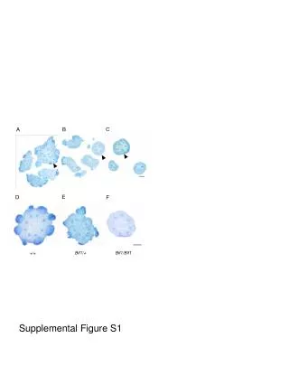
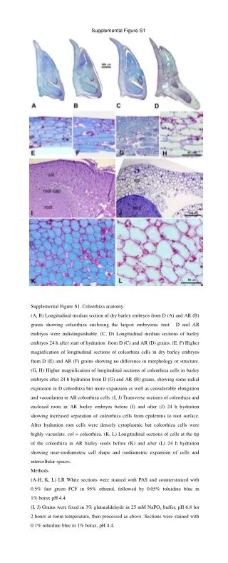
Supplemental Figure S1. Supplemental Figure S1. Coleorhiza anatomy.

E N D
Supplemental Figure S1 Supplemental Figure S1. Coleorhiza anatomy. (A, B) Longitudinal median section of dry barley embryos from D (A) and AR (B) grains showing coleorhiza enclosing the largest embryonic root. D and AR embryos were indistinguishable. (C, D) Longitudinal median sections of barley embryos 24 h after start of hydration from D (C) and AR (D) grains. (E, F) Higher magnification of longitudinal sections of coleorhiza cells in dry barley embryos from D (E) and AR (F) grains showing no difference in morphology or structure. (G, H) Higher magnification of longitudinal sections of coleorhiza cells in barley embryos after 24 h hydration from D (G) and AR (H) grains, showing some radial expansion in D coleorhiza but more expansion as well as considerable elongation and vacuolation in AR coleorhiza cells. (I, J) Transverse sections of coleorhiza and enclosed roots in AR barley embryos before (I) and after (J) 24 h hydration showing increased separation of coleorhiza cells from epidermis to root surface. After hydration root cells were densely cytoplasmic but coleorhiza cells were highly vacuolate. col = coleorhiza. (K, L) Longitudinal sections of cells at the tip of the coleorhiza in AR barley seeds before (K) and after (L) 24 h hydration showing near-isodiametric cell shape and isodiametric expansion of cells and intercellular spaces. Methods (A-H, K, L) LR White sections were stained with PAS and counterstained with 0.5% fast green FCF in 95% ethanol, followed by 0.05% toluidine blue in 1% borax pH 4.4. (I, J) Grains were fixed in 3% glutaraldehyde in 25 mM NaPO4 buffer, pH 6.8 for 2 hours at room temperature, then processed as above. Sections were stained with 0.1% toluidine blue in 1% borax, pH 4.4.