Download
1 / 71
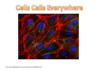
710 likes | 874 Vues
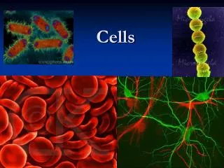
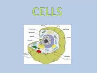
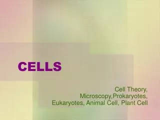
CELLS. Cell Theory, Microscopy,Prokaryotes, Eukaryotes, Animal Cell, Plant Cell. CELL THEORY. All living things are composed of cells and their products. New cells are formed only by the division of existing cells.

E N D
CELLS Cell Theory, Microscopy,Prokaryotes, Eukaryotes, Animal Cell, Plant Cell
CELL THEORY • All living things are composed of cells and their products. • New cells are formed only by the division of existing cells. • The cell is the functioning unit of life; the chemical reactions of life take place within cells.
Compound light microscopes Use visible light and a combination of lenses to magnify objects up to 1000 times.
Electron microscopes Use a beam of electrons, instead of light, to produce image
Transmission Electron Microscope (TEM) • Extremely thin sections • Electrons pass through some parts of specimen and not others, forming image • Good for organelle structure • Magnifies up to 250,000 times
1 µm Scanning electron microscopy (SEM) Cilia LE 6-4 Longitudinal section of cilium Transmission electron microscopy (TEM) Cross section of cilium 1 µm
Scanning Electron Microscope • Scans sample with a beam of electrons. • Magnifies up to 100,000 times
Advantages of light microscopes • Easy, inexpensive sample preparation • Allows examination of live material (movement; no artificial structures) • Colors can be seen (natural and stains) • Field of view is relatively large (1.8 mm at 100X magnification)
Advantage of electron microscopes • Excellent resolution, allowing for extremely high magnification • This permits examination of very small objects and details of cell structure
Prokaryotic Cells (Bacteria) • Pro – before • Karyon – nucleus Prokaryote = Before Nucleus
The outer layer has 2 parts • Cell wall – forms protective outer layer which prevents damage from outside and bursting if internal pressure is high • Plasma membrane – controls entry and exit of substances, some by active transport
CELLS Eukaryotic Cells
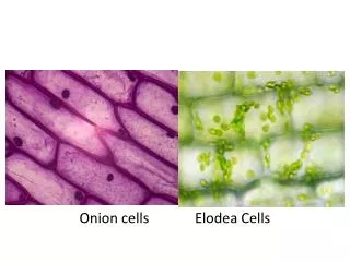
Prokaryotic vs. Eukaryotic Cells • Cells are prokaryotic or eukaryotic • Prokaryotic cells are Bacteria • Protozoa, Fungi, Animals, and Plants all consist of eukaryotic cells
Prokaryotic vs Eukaryotic Cells • Similarities • Plasma membrane • Semifluid substance called the cytoplasm • DNA - Chromosomes (carry genes) • Ribosomes (make protein) Differences • Prokaryotic cells have no nucleus • Prokaryotic cells lack membrane-bound organelles • Eukaryotic cells have DNA in a nucleus that is bound by a membrane (nuclear envelope) • Eukaryotic cells have membrane-bound organelles • Organelle: one of several formed bodies with specialized functions, suspended in the cytoplasm of eukaryotic cells
Surface area increases while Total volume remains constant 5 1 1 Total surface area (height x width x number of sides x number of boxes) LE 6-7 750 6 150 Total volume (height x width x length X number of boxes) 125 125 1 Surface-to-volume ratio (surface area volume) 6 6 1.2
Plasma membrane must have sufficient surface area to service the volume of the cell • The plasma membrane is a selective barrier that allows passage of oxygen, nutrients, and waste
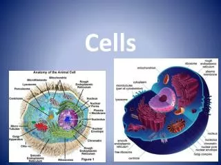
A Panoramic View of the Eukaryotic Cell • A eukaryotic cell has internal membranes that partition the cell into organelles • Plant and animal cells have most of the same organelles
ENDOPLASMIC RETICULUM (ER Nuclear envelope Flagellum Rough ER Smooth ER NUCLEUS Nucleolus Chromatin Centrosome Plasma membrane CYTOSKELETON Microfilaments LE 6-9a Intermediate filaments Microtubules Ribosomes: Microvilli Golgi apparatus • In animal cells but not plant cells: • Lysosomes • Centrioles • Flagella (in some plant sperm) Peroxisome Mitochondrion Lysosome
Nuclear envelope Rough endoplasmic reticulum NUCLEUS Nucleolus Chromatin Smooth endoplasmic reticulum Centrosome Ribosomes (small brown dots) Central vacuole Golgi apparatus Microfilaments Intermediate filaments CYTOSKELETON LE 6-9b Microtubules Mitochondrion Peroxisome Chloroplast Plasma membrane • In plant cells but not animal cells: • Chloroplasts • Central vacuole • Cell wall • Plasmodesmata Cell wall Plasmodesmata Wall of adjacent cell
Proteoglycan complex EXTRACELLULAR FLUID Collagen fiber Extra-Cellular Matrix Fibronectin LE 6-29a Plasma membrane CYTOPLASM Micro- filaments Integrin
Membranes Structure and Function
Plasma Membrane • Also known as “cell membrane” • Surrounds all cells • In cells with cell walls, the plasma membrane is found inside the cell wall
Phospholipid molecules have a hydrophilic region and a hydrophobic region
Most cells have watery environment on both sides of membrane • Water attracts the polar phosphate ends of the phospholipids • Phospholipids align to form double layer membrane, with polar ends on outside of each layer of the membrane • Non-polar tails are inside the bilayer LE 7-2 WATER Hydrophilic head Hydrophobic tail WATER
Hydrophilic region of protein LE 7-3 Phospholipid bilayer Hydrophobic region of protein
Extracellular layer Proteins Knife LE 7-4 Plasma membrane Cytoplasmic layer Extracellular layer Cytoplasmic layer
Membrane Proteins • A membrane is a collage of different proteins embedded in the fluid matrix of the lipid bilayer • Peripheral proteins are not embedded, they are attached to the membrane surface • Integral proteins penetrate the hydrophobic core and often span the membrane
Fibers of extracellular matrix (ECM) Glycoprotein Carbohydrate Glycolipid EXTRACELLULAR SIDE OF MEMBRANE LE 7-7 Cholesterol Peripheral proteins Microfilaments of cytoskeleton Integral protein CYTOPLASMIC SIDE OF MEMBRANE
LE 7-5c Cholesterol Cholesterol within the animal plasma membrane
Functions of Membrane Proteins • Proteins determine most of the membrane’s functions. They serve as: • Hormone binding sites • Enzymes • Cell–Cell joining & Communication • Channels for passive transport • Pumps for active transport
Transport across the plasma membrane Remember….. The plasma membrane controls what comes in and out of the cell.
Selective Permeability • Most biologic membranes are selectively or semi-permeable • This means that they allow some things through, but not others
Permeability • If substance CAN diffuse across membrane, membrane is permeable to that substance • If substance CANNOT diffuse across membrane, membrane is impermeable to the substance
One Way that Some Substances can Cross the Membrane is by Diffusion • Definition of diffusion: • Movement of particles from area where they are more concentrated to area where they are less concentrated
Diffusion, cont’d. • Diffusion is the tendency for molecules to spread out evenly into the available space. • Substances diffuse down their concentration gradient, that is, from an area where they are more highly concentrated to an area where they are less concentrated.
Molecules of dye Membrane (cross section) WATER LE 7-11a Net diffusion Net diffusion Equilibrium Diffusion of one solute
LE 7-11b Net diffusion Net diffusion Equilibrium Equilibrium Net diffusion Net diffusion Diffusion of two solutes
Diffusion, cont’d. • Diffusion across plasma membrane is a form of passive transport, because no work must be done to move substances down the concentration gradient • Passive Transport: transport across the membrane that requires no energy from the cell.
Diffusion, cont’d. • Hydrophobic, non-polar molecules can dissolve in and cross a membrane unassisted. • Hydrocarbons • CO2 • O2 Animation: Diffusion
Facilitated Diffusion • Diffusion of some other substances across the cell membrane is assisted, or “facilitated,” by protein channels within the membrane • Usually involves large or strongly charged molecules, which cannot dissolve in the lipid bilayer.
Facilitated diffusion • Even though movement is facilitated, it will still only occur from region of high concentration to region of low concentration. • Facilitated diffusion is still PASSIVE transport
Examples of molecules moving via facilitated diffusion • Some ions and polar molecules (e.g. water) • Aquaporins are channel proteins which greatly speed up the diffusion of water. Animation: Membrane Selectivity