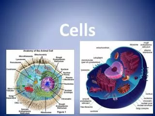
Cells
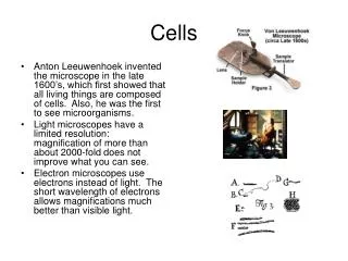
Cells. Anton Leeuwenhoek invented the microscope in the late 1600’s, which first showed that all living things are composed of cells. Also, he was the first to see microorganisms.

Cells
E N D
Presentation Transcript
Cells • Anton Leeuwenhoek invented the microscope in the late 1600’s, which first showed that all living things are composed of cells. Also, he was the first to see microorganisms. • Light microscopes have a limited resolution: magnification of more than about 2000-fold does not improve what you can see. • Electron microscopes use electrons instead of light. The short wavelength of electrons allows magnifications much better than visible light.
The Cell Theory • Use of the microscope for 150 years or so led to these basic beliefs about cells: • 1. All living things are made of cells. • 2. The cell is the smallest unit of life. • 3. All cells arise from pre-existing cells.
Basic Cell Organization • All cells contain: • 1. cell membrane that keeps the inside and outside separate. • 2. DNA-containing region that holds the instructions to run the processes of life. • 3. Cytoplasm: a semi-fluid region containing the rest of the cell’s machinery. • Prokaryotes: (bacteria): simple cells with no internal membrane-bound structures. DNA is in a special region of the cytoplasm. • Eukaryotes: complex cells with internal membranes. DNA is in a nucleus separated from the cytoplasm by a membrane.
Why Cells? • The basic problem is surface-to-volume ratio. All food and oxygen has to come in through the cell’s surface. As size increases, you get less surface area to support a given volume of cell contents. • For example, if the cell’s diameter increases: 1-2-3-4-5, its surface area increases 1-4-9-16-25, and its volume increases 1-8-27-64-125. A 5-fold increase in diameter cuts the amount of surface area per volume to 1/5 of the original: the cell starves. • Also, consider how a cell responds to a change in the environment: the signal must travel from the surface of the cell to the nucleus, then the nucleus issues new instructions to deal with the situation. The instructions must reach all parts of the cell. The bigger the cell, the longer it takes to respond to the environment. • Thus, cells are limited to small sizes, and multicellular organisms are composed of many cells. • Some minor exceptions: long thin cells, like nerve cells, can be several feet long. Also, cells can increase their surface area by using frilly membranes, like the cells that absorb food in the small intestine.
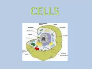
Eukaryotic Cells • Eukaryotic cells contain internal membranes and organelles. An organelle is an internal membrane bound structure that serves some specialized function within the cell. • Organelles we will discuss: • Cell membrane • Nucleus • Cytomembrane system, including endoplasmic reticulum, Golgi apparatus, vesicles, lysosomes, and peroxisomes • Mitochondria • Cytoskeleton • Special plant organelles: chloroplast, central vacuole, cell wall
Cell Membrane • Composed of phospholipids, with a polar (and therefore hydrophilic) head group, and 2 non-polar (hydrophobic) tails. A bilayer with the polar heads on the outsides and hydrophobic tails inside satisfies all of the molecule. The membrane is a “phospholipid bilayer”. • The membrane also contains cholesterol and various proteins. The proteins act as sensors, attachment points, cell recognition, or they transport small molecules through the membrane. • Membrane proteins and membrane lipids often have sugars attached to their outside edges: glycoproteins and glycolipids. For example, the differences between the ABO blood groups are due to differences in sugars attached to the outer membranes of red blood cells.
Cell Membrane, pt. 2 • The molecules in the membrane can move about like ships floating on the sea: the membrane is a two-dimensional fluid • In some cells, the membrane proteins are held in fixed positions by a network of proteins just under the membrane, a cytoskeleton. • Only water, a few gasses, and a few other small non-polar molecules can move freely through a pure phospholipid membrane. Everything else must be transported into the cell by protein channels in the membrane.
Transport Across the Cell Membrane • Basic rule: things spontaneously move from high concentration to low concentration (downhill). This process is called diffusion. • To get things to move from low to high (uphill), you need to add energy. In the cell, energy is kept in the form of ATP. • Three basic transport mechanisms: passive transport for downhill, active transport for uphill, and bulk transport for large amounts of material in either direction. • Also need to deal with excess water entering the cell.
Passive and Active Transport • Passive transport uses protein channels through the membrane that allow a particular molecule to go through it, down the concentration gradient. The speed and direction of movement depends on the relative concentrations inside and outside. Glucose is a good example: since cells burn glucose for energy, the concentration inside is less than the concentration outside. • Active transport uses proteins as pumps to concentrate molecules against the concentration gradient. The pumps use ATP for energy. One example is the calcium pump, which keeps the level of calcium ions in the cell 1000 times lower than outside, by constantly pumping calcium ions out. The balance of sodium and potassium ions is maintained with potassium high inside and sodium low inside, using a pump. Up to 1/3 of all energy used by the cell goes into maintaining the sodium/potassium balance.
Bulk Transport • In bulk transport, materials can move into the cell (called endocytosis) or out of the cell (called exocytosis). The two processes are reverses of each other. • In endocytosis, an area of the cell membrane forms an indentation that gradually pinches off into a small, self-contained membrane-bound sphere called a vesicle. The vesicle contains material that used to be outside the cell. An example is white blood cells engulfing and killing bacteria that have invaded the body. • In exocytosis, material the cell wishes to remove is contained in a vesicle. The vesicle fuses with the cell membrane, releasing the contents to the outside world. This is the way in which digestive enzymes are released into the stomach.
Water in the Cell • Water also moves down the concentration gradient. It moves into the cell to dilute the many molecules that are concentrated there. This process is called osmosis, and it exerts a pressure that can cause cells to swell up and burst. We say that pure water is hypotonic relative to the inside of a cell: pure water has fewer particles in it, so the water moves into the cell. • Conversely, if cells are put into a concentrated salt solution, water will leave the cells, moving to dilute the water outside. The concentrated salt solution is hypertonic: has more particles in it than the inside of the cell. • Normal body fluids are isotonic, having the same concentration of particles as the inside of the cell. • Cells need to defend themselves from the bad effects of osmosis, by keeping the concentration of water constant inside the cell.
Response to Osmotic Pressure • Plants have a simple defense against osmotic pressure: their cells are enclosed in a rigid cell wall, which resist the pressure so the cells don’t burst. If plants get too dry, they wilt because their cells are no longer held rigidly against the cell walls by osmotic pressure. Fungi and bacteria also use cell walls. • Many protists that live in fresh water have a special organelle that constantly pumps pure water out of the cell. • Animal cells constantly pump various ions (mostly sodium and potassium) in and out of the cell to combat osmotic pressure.
Nucleus • The nucleus issues instructions to build and maintain the cell, respond to changes in the environment, and to divide into 2 cells. • The cell’s instructions are coded in the DNA, which is the main part of chromosomes. A chromosome is composed of a single DNA molecule plus the proteins that support it and control it. • Most eukaryotes have a small number of chromosomes: humans have 46 chromosomes, corn plants have 20. The number is fixed within a species: all humans have 46 chromosomes except for some genetic oddities. • Each instruction in the DNA is called a gene. The genes issue their instructions, get expressed, as RNA copies. AN RNA copy of a gene is called messenger RNA (mRNA). The mRNA instructions move out of the membrane into the cytoplasm, where they are translated into proteins. • The translation of RNA messages into proteins is accomplished by ribosomes, which are structures made of both RNA and protein.
Nucleus, pt. 2 • Ribosomes are made in a special part of the nucleus, called the nucleolus. • However, the translation of messenger RNA into proteins by the ribosomes occurs in the cytoplasm outside the nucleus. Both the ribosomes and the messages move out of the nucleus into the cytoplasm to function. • The nucleus is surrounded by a double membrane called the nuclear envelope. It is studded with pores (made of protein) that let the ribosomes and the RNA messages out into the cytoplasm.
Cytomembrane System • The cytomembrane system is a group of organelles that has 3 basic functions: to manufacture new lipids and membranes, to modify polypeptides into their final proteins, and to synthesize and package proteins and other molecules for export. • We will talk about 4 organelles as part of this system: the endoplasmic reticulum (ER), the Golgi bodies, the lysosomes, and the peroxisomes.
Endoplasmic Reticulum • “Reticulum” means network; the ER is a network of tubules in the cytoplasm, composed of membranes just like the cell membrane. It provides a membrane channel from the nucleus to the cell membrane. • Two types, connected together: rough ER and smooth ER • Rough ER looks rough because it is studded with ribosomes, the cellular machines that synthesize proteins. Ribosomes on the rough ER make the proteins that go into the membrane, using the instructions from messenger RNA. Other ribosomes, not attached to the ER, make other proteins. • Smooth ER has no ribosomes. It is used to synthesize the lipids of the membrane. It is also used in liver cells to detoxify harmful chemicals in the blood. Other functions as well.
Golgi Body and Secretion • Proteins that are synthesized in the rough ER get finished in the Golgi body: sugars and phosphates added. • Golgi looks like a series of stacked plates. • Vesicles carry proteins from the ER to the Golgi, and then from the Golgi body to the cell membrane. Secretion to the outside world occurs by exocytosis: the vesicle fuses with the cell membrane, releasing its contents. • Proteins synthesized into the membrane of the ER end up in the cell membrane by the same mechanism • Basic mechanism of secretion: • genes are copied into messenger RNA in the nucleus • mRNA leaves the nucleus and attaches to ribosomes in the cytoplasm. • the ribosomes move to the rough ER and synthesize new proteins • proteins are transported by vesicles to the Golgi for finishing • proteins are transported in other vesicles to the cell membrane, where they are released from the cell.
Lysosomes and Peroxisomes • Lysosomes are intracellular stomachs: they are full of digestive enzymes that operate at low pH. You can think of them as little acid vats. Vesicles transport materials to the lysosomes, and the lysosomes digest them. In the process of “programmed cell death”, cells scheduled to die are destroyed from within by their lysosomes. An example is the tail of a tadpole, which is destroyed to make a tailless frog. • Lysosomal storage diseases are caused by genetic defects. An example is Gaucher disease, in which certain lipids accumulate inside of lysosomes instead of being broken down. This leads to interference with bone marrow function: blood and bone problems. • Peroxisomes are membrane-bound sacs used to break down fatty acids and some other molecules. They generate hydrogen peroxide, a poisonous molecule, in the process, which is the source of the name peroxisome.
Mitochondria • The mitochondria are the site where most of the cell’s ATP is generated, when organic compounds are broken down to carbon dioxide and water, using oxygen. • All eukaryotes have mitochondria. The number in a cell depends on that cell’s energy needs. • Mitochondria have their own circular DNA, the same kind found in bacteria. This and other evidence has led to the theory that mitochondria were once free-living bacteria that developed a mutually beneficial relationship with a primitive eukaryotic cell. • Mitochondria have 2 membranes, forming 2 compartments inside. To generate energy, hydrogen ions are accumulated between the 2 membranes. Then they flow down the concentration gradient into the inner compartment through a protein that uses the energy of their flow to create ATP. • Genetic defects in the mitochondria affect tissues that use a lot of energy: nerves, muscles, liver, kidney. They are unusual because they are inherited strictly from the mother—only the egg’s mitochondria go into the next generation.
Cytoskeleton • The cytoskeleton consists of proteins that give the cell shape. Without the cytoskeleton, cells would all be spherical. The cytoskeleton also causes organelles to move within the cell, and causes the cells themselves to move about. • The three main components of the cytoskeleton are: microtubules, microfilaments, and intermediate filaments, • Microtubules are long hollow tubes made up of many subunits called tubulin. They are used to pull the chromosomes apart during cell division, and to transport vesicles around inside the cell. • Microtubules can also be put into a special circular arrangement to form cilia and flagella. Special “motor proteins” cause these structures to have a whip-like motion, which propels the cell: a human sperm cell has a flagellum as a tail.
Cytoskeleton, pt. 2 • Microfilaments are composed of actin, which is also used in muscles. Organelles move inside the cell by using a motor protein (myosin) to pull themselves along the actin microfilaments. • The rapid assembly and disassembly of microfilaments just under the cell’s surface causes the movements of amoebas and various animal cells • Intermediate filaments are composed of several different proteins. They strengthen cells and give them their basic shape.
More Cytoskeleton • There are many genetic diseases affecting the cytoskeleton proteins. • One of the worst is Duchenne muscular dystrophy (DMD). This disease causes the muscles to swell up and eventually lose all strength, with the victims in wheelchairs by age 12 and dead of respiratory failure by age 20. • DMD is caused by the failure of the protein dystrophin to hold the actin microfilaments inside the muscle cells to their attachment points on the membrane. When the cells move, they rip their membranes apart and die. The muscle cells swell and burst as fluid enters through the torn membranes.
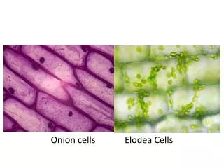
Plant Cell Organelles • Plants have three special structures not found in animals: the chloroplast, the cell wall, and the central vacuole. • The chloroplast is the site of photosynthesis, the process of converting carbon dioxide into sugar and oxygen using sunlight. Most life depends on photosynthesis, directly or indirectly. It uses the green pigment chlorophyll to capture the energy from light. • Like the mitochondria, chloroplasts have two membranes and their own circular DNA. Chloroplasts are also thought to have originated from an ancient mutually beneficial relationship between photosynthetic bacteria and a primitive eukaryote. • In some plant cells, chloroplasts are modified to store starch (as in potatoes) or to contain other pigments (as in flowers).
More Plant Organelles • Each plant cell is surrounded by a rigid cell wall made of cellulose and polysaccharides. The cell wall is outside of the cell membrane. In woody plants, the cell walls can become very thick and rigid. • Plant cells contain a central vacuole, which stores water. Osmotic pressure from the central vacuole squeezes the rest of the cytoplasm against the cell wall, giving the cell its strength.
Prokaryotic Cells • No internal membranes or organelles. • DNA loose in the cytoplasm. • Has a cell membrane, surrounded by a rigid cell wall that gives it shape. • Sometimes also a polysaccharide capsule surrounding the cell wall. • Flagella used for propulsion. Different structure than eukaryotic flagella. • Not much internal structure, but prokaryotes have a very wide variety of internal metabolic systems, and they inhabit a much wider range of habitats than eukaryotes.