Download
1 / 5
50 likes | 356 Vues
Supplementary Figures 1-3 The protein tyrosine-phosphatase DEP-1/PTPRJ promotes breast cancer cell invasion and metastasis Kathleen Spring 1# , Patrick Fournier 1# , Line Lapointe 1 , Catherine Chabot 1 , Jacinthe Roussy 1 , Sandra Pommey 1 , John Stagg 1, 2 , and Isabelle Royal 1, 3

E N D
Supplementary Figures 1-3 The protein tyrosine-phosphatase DEP-1/PTPRJ promotes breast cancer cell invasion and metastasis Kathleen Spring1#, Patrick Fournier1#, Line Lapointe1, Catherine Chabot1, Jacinthe Roussy1, Sandra Pommey1, John Stagg1, 2, and Isabelle Royal1, 3 1CRCHUM - Centre de recherche du Centre Hospitalier de l'Université de Montréal, and Institut du Cancer de Montréal, Montréal, QC, Canada; 2Faculty of Pharmacy and 3Department of Medicine, Université de Montréal, Montréal, QC, Canada.
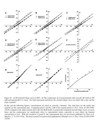
Supplementary Figure 1 - Spring et al. MDA-MB-231 Hs578T siCTL siCTL siDEP-1 siDEP-1 0’ 5’ 10’ 0’ 5’ 10’ 5% FBS 0’ 5’ 10’ 0’ 5’ 10’ 5% FBS DEP-1 DEP-1 pY418Src pY418 Src non-pY529Src non-pY529Src Src Src pY421Cortactin pY421Cortactin Cortactin Cortactin PLCγ PLCγ Supplementary Figure 1. DEP-1 promotes Src and Cortactin activation in invasive breast cancer cells.MDA-MB-231 and Hs578T cells transfected with control (CTL) or DEP-1 (#6; Dharmacon) siRNAs were stimulated with FBS (5%) for the indicated times. Activation of Src and its substrate Cortactin was evaluated by Western blotting on total cell lysates with phosphospecific antibodies. Results are representative of 3 independent experiments and confirm those obtained with the DEP-1 siRNA #3 from Qiagen (Fig. 2) and shRNAs (Fig. 4).
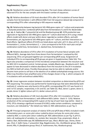
Supplementary Figure 2 - Spring et al. Supplementary Figure 2. Moderate expression of DEP-1 promotes Src activity in invasive breast cancer cells.MDA-MB-231 cells were transfected with empty vector (pmT2; 8 mg), or with increasing amounts of the WT DEP-1 plasmid (co-transfected with pmT2 for a constant amount of 8mg of DNA per condition). Cells were serum-starved and stimulated with FBS (5%) for the indicated times. Results show that moderately overexpressed DEP-1 (1 and 2 mg conditions) promoted Src Y529 dephosphorylation and Y418 phosphorylation. In contrast, higher expression of DEP-1 (3 mg and more; greater than a two-fold overexpression) led to its progressive incapacity to activate Src in MDA-MB-231 cells, as previously observed in endothelial cells (Spring et al. Blood, 2012). The average intensity of the DEP-1 immunoblot signals in cells overexpressing DEP-1 was plotted as the x-fold increase of DEP-1 expression over endogenous levels. *P < 0.05, **P < 0.01.
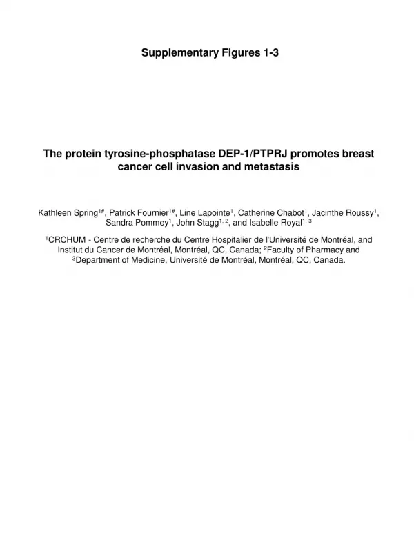
Supplementary Figure 3 - Spring et al. B A shScramble shDEP-1 2870 IHC: CD31 C shDEP-1 2870 shScramble 1 2 3 4 1 2 3 4 DEP-1 pY418Src non-pY529Src Src pY421Cortactin Cortactin PLCg D shScramble shDEP-1 2870 H&E
Supplementary Figure 3 - Spring et al. Supplementary Figure 3. Expression of DEP-1 in MDA-MB-231-derived tumors promotes the formation of lung metastases but not tumor growth or angiogenesis.(a) NSG mice were injected in the mammary fat pad with the shScramble or shDEP-1 2870 MDA-MD-231 cell populations. Tumor size was measured with calipers at the indicated time points (n=6 mice per group). (b) Paraffin-embedded tumor sections were stained for CD31 by IHC to monitor angiogenesis. Capillaries were counted per region of interest (n=6 mice per group). (c) Tumor cell lysates were immunoblotted with the indicated phosphospecific and expression level antibodies. The average phosphorylation levels of Src and Cortactin relative to their protein expression levels in the shDEP-1 tumors were determined by densitometry analysis and normalized to those of the control shScramble condition. The levels of DEP-1 were normalized to those of PLCg (n=4 mice per group). **P < 0.01. Decreased phosphorylation of Src Y418 and Cortactin Y421, as well as decreased dephosphorylation of Src Y529, are indicative of reduced Src signaling in the shDEP-1 2870 tumors. (d) Lungs cryosections were H&E stained and the average metastatic burden (%) was calculated (n=6 mice per group). *P < 0.05. These results demonstrate that depletion of DEP-1 with the #2870 shRNA yields results that follow the same trend as those obtained with the #2872 shRNA (Fig. 6), despite its reduced capacity to silence DEP-1 expression.