Crystal Uptake Inhibition Effects on MDCK Cell Viability and Adhesion
This study examines the impact of inhibitors on crystal uptake and cell viability in MDCK cells. Results show percentages of viable cells and numbers of adhered/internalized crystals after various treatments. Findings suggest potential strategies for inhibiting crystal uptake in cells.

Crystal Uptake Inhibition Effects on MDCK Cell Viability and Adhesion
E N D
Presentation Transcript
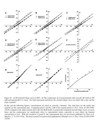
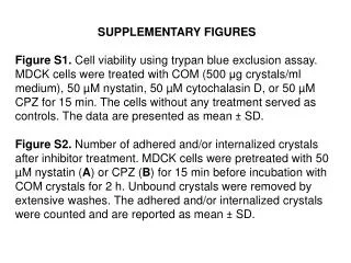
SUPPLEMENTARY FIGURES • Figure S1. Cell viability using trypan blue exclusion assay. MDCK cells were treated with COM (500 μg crystals/ml medium),50 µM nystatin, 50 µM cytochalasin D, or 50 µM CPZ for 15 min. The cells without any treatment served as controls. The data are presented as mean ± SD. • Figure S2.Number of adhered and/or internalized crystals after inhibitor treatment. MDCK cells were pretreated with 50 µM nystatin (A) or CPZ (B) for 15 min before incubation with COM crystals for 2 h. Unbound crystals were removed by extensive washes. The adhered and/or internalized crystals were counted and are reported as mean ± SD.
Supplementary Figure S1 Cell viability (%) (500 μg/ml) (50 μM) (50 μM) (50 μM)